Filter
Associated Lab
- Aso Lab (1) Apply Aso Lab filter
- Branson Lab (1) Apply Branson Lab filter
- Card Lab (4) Apply Card Lab filter
- Cardona Lab (17) Apply Cardona Lab filter
- Dickson Lab (1) Apply Dickson Lab filter
- Fetter Lab (9) Apply Fetter Lab filter
- Heberlein Lab (1) Apply Heberlein Lab filter
- Riddiford Lab (7) Apply Riddiford Lab filter
- Rubin Lab (4) Apply Rubin Lab filter
- Simpson Lab (2) Apply Simpson Lab filter
- Singer Lab (1) Apply Singer Lab filter
- Stern Lab (4) Apply Stern Lab filter
- Remove Truman Lab filter Truman Lab
- Zlatic Lab (13) Apply Zlatic Lab filter
Associated Project Team
Publication Date
- 2023 (2) Apply 2023 filter
- 2021 (3) Apply 2021 filter
- 2020 (3) Apply 2020 filter
- 2019 (4) Apply 2019 filter
- 2018 (8) Apply 2018 filter
- 2017 (6) Apply 2017 filter
- 2016 (11) Apply 2016 filter
- 2015 (6) Apply 2015 filter
- 2014 (1) Apply 2014 filter
- 2013 (3) Apply 2013 filter
- 2012 (3) Apply 2012 filter
- 2011 (1) Apply 2011 filter
- 2010 (4) Apply 2010 filter
- 2009 (2) Apply 2009 filter
- 2008 (1) Apply 2008 filter
Type of Publication
- Remove Janelia filter Janelia
58 Publications
Showing 31-40 of 58 resultsAnimals use sensory information to move toward more favorable conditions. Drosophila larvae can move up or down gradients of odors (chemotax), light (phototax), and temperature (thermotax) by modulating the probability, direction, and size of turns based on sensory input. Whether larvae can anemotax in gradients of mechanosensory cues is unknown. Further, although many of the sensory neurons that mediate taxis have been described, the central circuits are not well understood. Here, we used high-throughput, quantitative behavioral assays to demonstrate Drosophila larvae anemotax in gradients of wind speeds and to characterize the behavioral strategies involved. We found that larvae modulate the probability, direction, and size of turns to move away from higher wind speeds. This suggests that similar central decision-making mechanisms underlie taxis in somatosensory and other sensory modalities. By silencing the activity of single or very few neuron types in a behavioral screen, we found two sensory (chordotonal and multidendritic class III) and six nerve cord neuron types involved in anemotaxis. We reconstructed the identified neurons in an electron microscopy volume that spans the entire larval nervous system and found they received direct input from the mechanosensory neurons or from each other. In this way, we identified local interneurons and first- and second-order subesophageal zone (SEZ) and brain projection neurons. Finally, silencing a dopaminergic brain neuron type impairs anemotaxis. These findings suggest that anemotaxis involves both nerve cord and brain circuits. The candidate neurons and circuitry identified in our study provide a basis for future detailed mechanistic understanding of the circuit principles of anemotaxis.
Small animals navigate in the environment as a function of varying sensory information in order to reach more favorable environmental conditions. To achieve this Drosophila larvae alternate periods of runs and turns in gradients of light, temperature, odors and CO2. While the sensory neurons that mediate the navigation behaviors in the different sensory gradients have been described, where and how are these navigational strategies are implemented in the central nervous system and controlled by neuronal circuit elements is not well known. Here we characterize for the first time the navigational strategies of Drosophila larvae in gradients of air-current speeds using high-throughput behavioral assays and quantitative behavioral analysis. We find that larvae extend runs when facing favorable conditions and increase turn rate when facing unfavorable direction, a strategy they use in other sensory modalities as well. By silencing the activity of individual neurons and very sparse expression patterns (2 or 3 neuron types), we further identify the sensory neurons and circuit elements in the ventral nerve cord and brain of the larva required for navigational decisions during anemotaxis. The phenotypes of these central neurons are consistent with a mechanism where the increase of the turning rate in unfavorable conditions and decrease in turning rate in favorable conditions are independently controlled.
Drosophila central neurons arise from neuroblasts that generate neurons in a pair-wise fashion, with the two daughters providing the basis for distinct A and B hemilineage groups. Thirty three postembryonically-born hemilineages contribute over 90% of the neurons in each thoracic hemisegment. We devised genetic approaches to define the anatomy of most of these hemilineages and to assessed their functional roles using the heat-sensitive channel dTRPA1. The simplest hemilineages contained local interneurons and their activation caused tonic or phasic leg movements lacking interlimb coordination. The next level was hemilineages of similar projection cells that drove intersegmentally coordinated behaviors such as walking. The highest level involved hemilineages whose activation elicited complex behaviors such as takeoff. These activation phenotypes indicate that the hemilineages vary in their behavioral roles with some contributing to local networks for sensorimotor processing and others having higher order functions of coordinating these local networks into complex behavior.
The vast majority of the adult fly ventral nerve cord is composed of 34 hemilineages, which are clusters of lineally related neurons. Neurons in these hemilineages use one of the three fast-acting neurotransmitters (acetylcholine, GABA, or glutamate) for communication. We generated a comprehensive neurotransmitter usage map for the entire ventral nerve cord. We did not find any cases of neurons using more than one neurotransmitter, but found that the acetylcholine specific gene ChAT is transcribed in many glutamatergic and GABAergic neurons, but these transcripts typically do not leave the nucleus and are not translated. Importantly, our work uncovered a simple rule: All neurons within a hemilineage use the same neurotransmitter. Thus, neurotransmitter identity is acquired at the stem cell level. Our detailed transmitter- usage/lineage identity map will be a great resource for studying the developmental basis of behavior and deciphering how neuronal circuits function to regulate behavior.
Visual systems transduce, process and transmit light-dependent environmental cues. Computation of visual features depends on photoreceptor neuron types (PR) present, organization of the eye and wiring of the underlying neural circuit. Here, we describe the circuit architecture of the visual system of Drosophila larvae by mapping the synaptic wiring diagram and neurotransmitters. By contacting different targets, the two larval PR-subtypes create two converging pathways potentially underlying the computation of ambient light intensity and temporal light changes already within this first visual processing center. Locally processed visual information then signals via dedicated projection interneurons to higher brain areas including the lateral horn and mushroom body. The stratified structure of the larval optic neuropil (LON) suggests common organizational principles with the adult fly and vertebrate visual systems. The complete synaptic wiring diagram of the LON paves the way to understanding how circuits with reduced numerical complexity control wide ranges of behaviors.
During larval life most of the thoracic neuroblasts (NBs) in Drosophila undergo a second phase of neurogenesis to generate adult-specific neurons that remain in an immature, developmentally stalled state until pupation. Using a combination of MARCM and immunostaining with a neurotactin antibody Truman et al. (2004) identified 24 adult specific NB lineages within each thoracic hemineuromere of the larval ventral nervous system (VNS) but because the neurotactin labeling of lineage tracts disappearing early in metamorphosis they were unable extend the identification of the these lineages into the adult. Here we show that immunostaining with an antibody against the cell adhesion molecule Neuroglian reveals the same larval secondary lineage projections through metamorphosis and by identifying each neuroglian positive tract at selected stages we have traced the larval hemilineage tracts for all three thoracic neuromeres through metamorphosis into the adult. To validate tract identifications we used the genetic toolkit developed by Harris et al. (2015) to preserve hemilineage specific GAL4 expression patterns from larval into the adult stage. The immortalized expression proved a powerful confirmation of the analysis of the neuroglian scaffold. This work has enabled us to directly link the secondary, larval NB lineages to their adult counterparts. The data provide an anatomical framework that 1) makes it possible to assign most neurons to their parent lineage and 2) allows more precise definitions of the neuronal organization of the adult VNS based in developmental units/rules. This article is protected by copyright. All rights reserved.
Dopaminergic neurons (DANs) drive learning across the animal kingdom, but the upstream circuits that regulate their activity and thereby learning remain poorly understood. We provide a synaptic-resolution connectome of the circuitry upstream of all DANs in a learning center, the mushroom body of Drosophila larva. We discover afferent sensory pathways and a large population of neurons that provide feedback from mushroom body output neurons and link distinct memory systems (aversive and appetitive). We combine this with functional studies of DANs and their presynaptic partners and with comprehensive circuit modeling. We find that DANs compare convergent feedback from aversive and appetitive systems, which enables the computation of integrated predictions that may improve future learning. Computational modeling reveals that the discovered feedback motifs increase model flexibility and performance on learning tasks. Our study provides the most detailed view to date of biological circuit motifs that support associative learning.
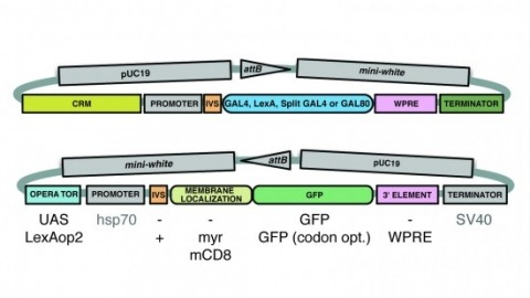
A wide variety of biological experiments rely on the ability to express an exogenous gene in a transgenic animal at a defined level and in a spatially and temporally controlled pattern. We describe major improvements of the methods available for achieving this objective in Drosophila melanogaster. We have systematically varied core promoters, UTRs, operator sequences, and transcriptional activating domains used to direct gene expression with the GAL4, LexA, and Split GAL4 transcription factors and the GAL80 transcriptional repressor. The use of site-specific integration allowed us to make quantitative comparisons between different constructs inserted at the same genomic location. We also characterized a set of PhiC31 integration sites for their ability to support transgene expression of both drivers and responders in the nervous system. The increased strength and reliability of these optimized reagents overcome many of the previous limitations of these methods and will facilitate genetic manipulations of greater complexity and sophistication.
Animal locomotion requires spatiotemporally coordinated contraction of muscles throughout the body. Here, we investigate how contractions of antagonistic groups of muscles are intersegmentally coordinated during bidirectional crawling of Drosophila larvae. We identify two pairs of higher-order premotor excitatory interneurons present in each abdominal neuromere that intersegmentally provide feedback to the adjacent neuromere during motor propagation. The two feedback neuron pairs are differentially active during either forward or backward locomotion but commonly target a group of premotor interneurons that together provide excitatory inputs to transverse muscles and inhibitory inputs to the antagonistic longitudinal muscles. Inhibition of either feedback neuron pair compromises contraction of transverse muscles in a direction-specific manner. Our results suggest that the intersegmental feedback neurons coordinate contraction of synergistic muscles by acting as delay circuits representing the phase lag between segments. The identified circuit architecture also shows how bidirectional motor networks could be economically embedded in the nervous system.
Multiple studies have investigated the mechanisms of aggressive behavior in Drosophila; however, little is known about the effects of chronic fighting experience. Here, we investigated if repeated fighting encounters would induce an internal state that could affect the expression of subsequent behavior. We trained wild-type males to become winners or losers by repeatedly pairing them with hypoaggressive or hyperaggressive opponents, respectively. As described previously, we observed that chronic losers tend to lose subsequent fights, while chronic winners tend to win them. Olfactory conditioning experiments showed that winning is perceived as rewarding, while losing is perceived as aversive. Moreover, the effect of chronic fighting experience generalized to other behaviors, such as gap-crossing and courtship. We propose that in response to repeatedly winning or losing aggressive encounters, male flies form an internal state that displays persistence and generalization; fight outcomes can also have positive or negative valence. Furthermore, we show that the activities of the PPL1-γ1pedc dopaminergic neuron and the MBON-γ1pedc>α/β mushroom body output neuron are required for aversion to an olfactory cue associated with losing fights.