Filter
Publication Date
Type of Publication
2 Publications
Showing 1-2 of 2 resultsThe antennae of male silk moths are extremely sensitive to the female sex pheromone such that a male moth can find a female up to 4.5 km away. This remarkable sensitivity is due to both the morphological and biochemical design of these antennae. Along the branches of the plumose antennae are the sensilla trichodea, each consisting of a hollow cuticular hair containing two unbranched dendrites bathed in a fluid, the receptor lymph ,3. The dendrites and receptor lymph are isolated from the haemolymph by a barrier of epidermal cells which secreted the cuticular hair. Pheromone molecules are thought to diffuse down 100 A-wide pore tubules through the cuticular wall and across the receptor lymph space to receptors located in the dendritic membrane. To prevent the accumulation of residual stimulant and hence sensory adaptation, the pheromone molecules are subsequently inactivated in an apparent two-step process of rapid ’early inactivation’ followed by much slower enzymatic degradation. The biochemistry involved in this sequence of events is largely unknown. We report here the identification of three proteins which interact with the pheromone of the wild silk moth Antheraea polyphemus: a pheromone-binding protein and a pheromone-degrading esterase, both uniquely located in the pheromone-sensitive sensilla; and a second esterase common to all cuticular tissues except the sensilla.
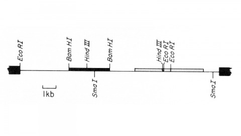
We describe the isolation of a cloned DNA segment carrying unique sequences from the white locus of Drosophila melanogaster. Sequences within the cloned segment are shown to hybridize in situ to the white locus region on the polytene chromosomes of both wild-type strains and strains carrying chromosomal rearrangements whose breakpoints bracket the white locus. We further show that two small deficiency mutations, deleting white locus genetic elements but not those of complementation groups contiguous to white, delete the genomic sequences corresponding to a portion of the cloned segment. The strategy we have employed to isolate this cloned segment exploits the existence of an allele at the white locus containing a copy of a previously cloned transposable, reiterated DNA sequence element. We describe a simple, rapid method for retrieving cloned segments carrying a copy of the transposable element together with contiguous sequences corresponding to this allele. The strategy described is potentially general and we discuss its application to the cloning of the DNA sequences of other genes in Drosophila, including those identified only by genetic analysis and for which no RNA product is known.