Filter
Associated Lab
- Druckmann Lab (1) Apply Druckmann Lab filter
- Grigorieff Lab (1) Apply Grigorieff Lab filter
- Heberlein Lab (1) Apply Heberlein Lab filter
- Lippincott-Schwartz Lab (2) Apply Lippincott-Schwartz Lab filter
- Magee Lab (1) Apply Magee Lab filter
- Menon Lab (1) Apply Menon Lab filter
- Pachitariu Lab (2) Apply Pachitariu Lab filter
- Spruston Lab (1) Apply Spruston Lab filter
- Stern Lab (1) Apply Stern Lab filter
- Sternson Lab (1) Apply Sternson Lab filter
- Turner Lab (1) Apply Turner Lab filter
Publication Date
- December 24, 2013 (1) Apply December 24, 2013 filter
- December 19, 2013 (1) Apply December 19, 2013 filter
- December 18, 2013 (2) Apply December 18, 2013 filter
- December 9, 2013 (1) Apply December 9, 2013 filter
- December 7, 2013 (1) Apply December 7, 2013 filter
- December 5, 2013 (4) Apply December 5, 2013 filter
- December 1, 2013 (3) Apply December 1, 2013 filter
- Remove December 2013 filter December 2013
- Remove 2013 filter 2013
Type of Publication
13 Publications
Showing 1-10 of 13 resultsThe gills of most teleost fishes are covered by plate-like structures, the secondary lamellae, that provide the bulk of the respiratory surface area. Water passing over the secondary lamellae exchanges gases with blood passing through the secondary lamellae, forming a system that has served as a classic model of counter-current exchange. In this study, a computational model of flow around the secondary lamellae is used to examine the hydrodynamic consequences of changes to the lamellar morphology. Consistent with previous studies, the interlamellar distance is found to strongly affect the hydrodynamic resistance of the gills. However, the presence of a small gap between the tips of the secondary lamellae is found to have a similarly strong effect on the hydrodynamic resistance and flow patterns within the gills. The results from this model have been generally formulated, allowing the calculation of the hydrodynamic resistance for measured morphometric parameters. These results provide a new basis for comparing theoretical predictions of the gill resistance with measured values, and provide a general model for examining the diversity gill morphologies observed in teleost fishes.
Neuronal computation involves the integration of synaptic inputs that are often distributed over expansive dendritic trees, suggesting the need for compensatory mechanisms that enable spatially disparate synapses to influence neuronal output. In hippocampal CA1 pyramidal neurons, such mechanisms have indeed been reported, which normalize either the ability of distributed synapses to drive action potential initiation in the axon or their ability to drive dendritic spiking locally. Here we report that these mechanisms can coexist, through an elegant combination of distance-dependent regulation of synapse number and synaptic expression of AMPA and NMDA receptors. Together, these complementary gradients allow individual dendrites in both the apical and basal dendritic trees of hippocampal neurons to operate as facile computational subunits capable of supporting both global integration in the soma/axon and local integration in the dendrite.
The neural circuits that mediate behavioral choice evaluate and integrate information from the environment with internal demands and then initiate a behavioral response. Even circuits that support simple decisions remain poorly understood. In Drosophila melanogaster, oviposition on a substrate containing ethanol enhances fitness; however, little is known about the neural mechanisms mediating this important choice behavior. Here, we characterize the neural modulation of this simple choice and show that distinct subsets of dopaminergic neurons compete to either enhance or inhibit egg-laying preference for ethanol-containing food. Moreover, activity in α'β' neurons of the mushroom body and a subset of ellipsoid body ring neurons (R2) is required for this choice. We propose a model where competing dopaminergic systems modulate oviposition preference to adjust to changes in natural oviposition substrates.
Biological tissue is often composed of cells with similar morphologies replicated throughout large volumes and many biological applications rely on the accurate identification of these cells and their locations from image data. Here we develop a generative model that captures the regularities present in images composed of repeating elements of a few different types. Formally, the model can be described as convolutional sparse block coding. For inference we use a variant of convolutional matching pursuit adapted to block-based representations. We extend the K-SVD learning algorithm to subspaces by retaining several principal vectors from the SVD decomposition instead of just one. Good models with little cross-talk between subspaces can be obtained by learning the blocks incrementally. We perform extensive experiments on simulated images and the inference algorithm consistently recovers a large proportion of the cells with a small number of false positives. We fit the convolutional model to noisy GCaMP6 two-photon images of spiking neurons and to Nissl-stained slices of cortical tissue and show that it recovers cell body locations without supervision. The flexibility of the block-based representation is reflected in the variability of the recovered cell shapes.
The final cleavage event that terminates cell division, abscission of the small, dense intercellular bridge, has been particularly challenging to resolve. Here, we describe imaging innovations that helped answer long-standing questions about the mechanism of abscission. We further explain how computational modeling of high-resolution data was employed to test hypotheses and generate additional insights. We present the model that emerges from application of these complimentary approaches. Similar experimental strategies will undoubtedly reveal exciting details about other underresolved cellular structures.
Freshly isolated, depolarized rat hepatocytes can repolarize into bile canalicular networks when plated in collagen sandwich cultures. We studied the events underlying this repolarization process, focusing on how hepatocytes restore ATP synthesis and resupply biosynthetic precursors after the stress of being isolated from liver. We found that soon after being plated in collagen sandwich cultures, hepatocytes converted their mitochondria into highly fused networks. This occurred through a combination of upregulation of mitochondrial fusion proteins and downregulation of a mitochondrial fission protein. Mitochondria also became more active for oxidative phosphorylation, leading to overall increased ATP levels within cells. We further observed that autophagy was upregulated in the repolarizing hepatocytes. Boosted autophagy levels likely served to recycle cellular precursors, supplying building blocks for repolarization. Repolarizing hepatocytes also extensively degraded lipid droplets, whose fatty acids provide precursors for ?-oxidation to fuel oxidative phosphorylation in mitochondria. Thus, through coordination of mitochondrial fusion, autophagy, and lipid droplet consumption, depolarized hepatocytes are able to boost ATP synthesis and biosynthetic precursors to efficiently repolarize in collagen sandwich cultures.
In the olfactory system, sensory inputs are arranged in different glomerular channels, which respond in combinatorial ensembles to the various chemical features of an odor. We investigated where and how this combinatorial code is read out deeper in the brain. We exploited the unique morphology of neurons in the Drosophila mushroom body, which receive input on large dendritic claws. Imaging odor responses of these dendritic claws revealed that input channels with distinct odor tuning converge on individual mushroom body neurons. We determined how these inputs interact to drive the cell to spike threshold using intracellular recordings to examine mushroom body responses to optogenetically controlled input. Our results provide an elegant explanation for the characteristic selectivity of mushroom body neurons: these cells receive different types of input and require those inputs to be coactive to spike. These results establish the mushroom body as an important site of integration in the fly olfactory system.
Mapping mammalian synaptic connectivity has long been an important goal of neuroscientists since it is considered crucial for explaining human perception and behavior. Yet, despite enormous efforts, the overwhelming complexity of the neural circuitry and the lack of appropriate techniques to unravel it have limited the success of efforts to map connectivity. However, recent technological advances designed to overcome the limitations of conventional methods for connectivity mapping may bring about a turning point. Here, we address the promises and pitfalls of these new mapping technologies.
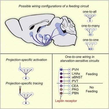
Neural circuits for essential natural behaviors are shaped by selective pressure to coordinate reliable execution of flexible goal-directed actions. However, the structural and functional organization of survival-oriented circuits is poorly understood due to exceptionally complex neuroanatomy. This is exemplified by AGRP neurons, which are a molecularly defined population that is sufficient to rapidly coordinate voracious food seeking and consumption behaviors. Here, we use cell-type-specific techniques for neural circuit manipulation and projection-specific anatomical analysis to examine the organization of this critical homeostatic circuit that regulates feeding. We show that AGRP neuronal circuits use a segregated, parallel, and redundant output configuration. AGRP neuron axon projections that target different brain regions originate from distinct subpopulations, several of which are sufficient to independently evoke feeding. The concerted anatomical and functional analysis of AGRP neuron projection populations reveals a constellation of core forebrain nodes, which are part of an extended circuit that mediates feeding behavior.
A new generation of direct electron detectors for transmission electron microscopy (TEM) promises significant improvement over previous detectors in terms of their modulation transfer function (MTF) and detective quantum efficiency (DQE). However, the performance of these new detectors needs to be carefully monitored in order to optimize imaging conditions and check for degradation over time. We have developed an easy-to-use software tool, FindDQE, to measure MTF and DQE of electron detectors using images of a microscope’s built-in beam stop. Using this software, we have determined the DQE curves of four direct electron detectors currently available: the Gatan K2 Summit, the FEI Falcon I and II, and the Direct Electron DE-12, under a variety of total dose and dose rate conditions. We have additionally measured the curves for the Gatan US4000 and TVIPS TemCam-F416 scintillator-based cameras. We compare the results from our new method with published curves.