Filter
Associated Lab
Publication Date
- November 1991 (2) Apply November 1991 filter
- October 1991 (1) Apply October 1991 filter
- September 1991 (1) Apply September 1991 filter
- July 1991 (2) Apply July 1991 filter
- May 1991 (2) Apply May 1991 filter
- March 1991 (2) Apply March 1991 filter
- January 1991 (1) Apply January 1991 filter
- Remove 1991 filter 1991
Type of Publication
11 Publications
Showing 1-10 of 11 resultsExamined kin discrimination and colony defense in soldier-producing aphids from the surface of 3 galls. Soldiers always attacked conspecific nonsoldiers, regardless of origin, and never attacked conspecific soldiers. Soldier attacks of nonsoldiers may exclude unrelated nonsoldier aphids from the gall where they would propagate and compete with resident aphids. (PsycINFO Database Record (c) 2012 APA, all rights reserved)

We have conducted a genetic screen for mutations that decrease the effectiveness of signaling by a protein tyrosine kinase, the product of the Drosophila melanogaster sevenless gene. These mutations define seven genes whose wild-type products may be required for signaling by sevenless. Four of the seven genes also appear to be essential for signaling by a second protein tyrosine kinase, the product of the Ellipse gene. The putative products of two of these seven genes have been identified. One encodes a ras protein. The other locus encodes a protein that is homologous to the S. cerevisiae CDC25 protein, an activator of guanine nucleotide exchange by ras proteins. These results suggest that the stimulation of ras protein activity is a key element in the signaling by sevenless and Ellipse and that this stimulation may be achieved by activating the exchange of GTP for bound GDP by the ras protein.
During a four month study of male territoriality males of the euglossine bee Eulaema meriana exhibited the two alternative behavior patterns of territoriality and transiency. Territorial males patrolled an area adjacent to a tree upon which they perched. Territorial males utilized the same territory for up to 49 days, though often not on consecutive days, and appeared to non-violently relinquish territories to new males. Transients did not defend territories but flew from one territory to another and flew with the territorial male around the territory, rarely bumping, and never grappling. Transient males left the territory soon after the territorial male flew back and forth in front of the perch tree in a zig-zag flight. The alternative behaviors were correlated with wing wear such that males with little wing wear defended territories and males with considerable wing wear pursued a transient strategy. Behavior patterns were not correlated with head width. Comparison of territory trees with the territory trees of a closely related species indicate that each species utilized trees of a certain diameter class for perching. In addition, analysis of hemispherical canopy photographs indicates that males appeared to prefer territories that received a maximum of diffuse sunlight but a minimum of direct sunlight. Both territorial and transient males consistently returned to specific territories over their lifetime but appeared to travel long distances to forage for fragrances. Territorial and transient males visited fragrance baits with equal frequency suggesting that non-territorial, as well as territorial, males required fragrances.
The maleless (mle) gene is one of four known regulatory loci required for increased transcription (dosage compensation) of X-linked genes in D. melanogaster males. A predicted mle protein (MLE) contains seven short segments that define a superfamily of known and putative RNA and DNA helicases. MLE, while present in the nuclei of both male and female cells, differs in its association with polytene X chromosomes in the two sexes. MLE is associated with hundreds of discrete sites along the length of the X chromosome in males and not in females. The predominant localization of MLE to the X chromosome in males makes it a strong candidate to be a direct regulator of dosage compensation.
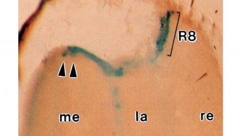
Histological staining of wild-type and sevenless transgenic Drosophila melanogaster bearing Rh3-lacZ fusion genes permits the selective visualization of polarization-sensitive R7 and R8 photoreceptor cells located along the dorsal anterior eye margin. Diffusion of beta-galactosidase throughout these cells reveals that they project long axons to the two most peripheral synaptic target rows of the dorsal posterior medulla, defining a specialized marginal zone of this optic lobe. Comparison of the staining patterns of marginal and nonmarginal Rh3-lacZ-expressing photoreceptor cells in the same histological preparations suggest that the marginal cells possess morphologically specialized axons and synaptic terminals. These findings are discussed with reference to the neuroanatomy of the corresponding dorsal marginal eye and optic lobe regions of the larger dipterans Musca and Calliphora, and in relation to the ability of Drosophila to orient to polarized light.
The expression of GABA is restricted to the progeny of only six of the 24 identified postembryonic lineages in the thoracic ganglia of the tobacco hornworm, Manduca sexta (Witten and Truman, 1991). It is colocalized with a peptide similar to molluscan small cardioactive peptide B (SCPB) in some of the neurons in two of the six lineages. By combining chemical ablation of the neuroblasts at specific larval stages with birth dating of the progeny, we tested whether the expression of GABA and the SCPB-like peptide was determined strictly by cell lineage or involved cellular interactions among the members of individual clonal groups. Chemical ablation of the six specific neuroblasts that produced the GABA-positive neurons (E, K, M, N, T, and X) or of the two that produced the GABA + SCPB-like-immunoreactive neurons (K, M) prior to the generation of their lineages resulted in the loss of these immunoreactivities. These results suggest that regulation between lineages did not occur. Ablation of the K and M neuroblasts after they had produced a small portion of their lineages had no effect on the expression of GABA, but did affect the pattern of the SCPB-like immunoreactivity. Combining birth-dating techniques with transmitter immunocytochemistry revealed that it was the position in the birth order and not interactions among the clonally related neurons that influenced the peptidergic phenotype. These results suggest that cell lineage is involved in establishing the GABAergic phenotype and that both cell lineage and birth order influence the determination of the peptidergic phenotype.(ABSTRACT TRUNCATED AT 250 WORDS)
Human mitochondrial transcription factor 1 (mtTF1) has been sequenced and is a nucleus-encoded DNA binding protein of 204 amino acids (24,400 daltons). Expression of human mtTF1 in bacteria yields a protein with correct physical properties and the ability to activate mitochondrial DNA promoters. Analysis of the protein’s sequence reveals no similarities to any other DNA binding proteins except for the existence of two domains that are characteristic of high mobility group (HMG) proteins. Human mtTF1 is most closely related to a DNA binding HMG-box region in hUBF, a human protein known to be important for transcription by RNA polymerase I.
Defects in mitochondrial DNA (mtDNA) are associated with several different human diseases, including the mitochondrial encephalomyopathies. The mutations include deletions but also duplications and point mutations. Individuals with MELAS (mitochondrial myopathy, encephalopathy, lactic acidosis and stroke-like episodes) carry a common A-to-G substitution in a highly conserved portion of the gene for transfer RNA(Leu(UUR)). Although the MELAS mutation may be comparable to the defect in the tRNA(Lys) gene associated with MERRF (myoclonus epilepsy associated with ragged-red fibres), it is also embedded in the middle of a tridecamer sequence necessary for the formation of the 3’ ends of 16S ribosomal RNA in vitro. We found that the MELAS mutation results in severe impairment of 16S rRNA transcription termination, which correlates with a reduced affinity of the partially purified termination protein for the MELAS template. This suggests that the molecular defect in MELAS is the inability to produce the correct type and quantity of rRNA relative to other mitochondrial gene products.
In near-field scanning optical microscopy, a light source or detector with dimensions less than the wavelength (lambda) is placed in close proximity (lambda/50) to a sample to generate images with resolution better than the diffraction limit. A near-field probe has been developed that yields a resolution of approximately 12 nm ( approximately lambda/43) and signals approximately 10(4)- to 10(6)-fold larger than those reported previously. In addition, image contrast is demonstrated to be highly polarization dependent. With these probes, near-field microscopy appears poised to fulfill its promise by combining the power of optical characterization methods with nanometric spatial resolution.
In near-field scanning optical microscopy, a light source or detector with dimensions less than the wavelength (lambda) is placed in close proximity (lambda/50) to a sample to generate images with resolution better than the diffraction limit. A near-field probe has been developed that yields a resolution of approximately 12 nm ( approximately lambda/43) and signals approximately 10(4)- to 10(6)-fold larger than those reported previously. In addition, image contrast is demonstrated to be highly polarization dependent. With these probes, near-field microscopy appears poised to fulfill its promise by combining the power of optical characterization methods with nanometric spatial resolution.
Commentary: Introduced the adiabatically tapered single mode fiber probe to near-field scanning optical microscopy which, together with shear force feedback, made the technique a practical reality. Although earlier claims of superresolution via near-field microscopy existed for nearly a decade, this paper was the first to convincingly break Abbe’s limit with visible light, as demonstrated by reproducibly resolving known, complex nanoscale patterns having features separated by much less than the wavelength. Whereas our fiber probe and shear force technologies were soon widely adopted and key to many novel applications (see above), the earlier methods proved to be technological dead ends, never achieving the results of their original claims. This experience taught me the most valuable lesson of my career: while it’s bad to bullshit others, it’s even worse to bullshit yourself. It’s a lesson sadly unheeded by many current practitioners of superresolution microscopy.