Filter
Associated Lab
- Aso Lab (1) Apply Aso Lab filter
- Betzig Lab (2) Apply Betzig Lab filter
- Egnor Lab (1) Apply Egnor Lab filter
- Grigorieff Lab (1) Apply Grigorieff Lab filter
- Jayaraman Lab (1) Apply Jayaraman Lab filter
- Lavis Lab (1) Apply Lavis Lab filter
- Liu (Zhe) Lab (1) Apply Liu (Zhe) Lab filter
- Rubin Lab (3) Apply Rubin Lab filter
- Saalfeld Lab (1) Apply Saalfeld Lab filter
- Singer Lab (2) Apply Singer Lab filter
- Spruston Lab (1) Apply Spruston Lab filter
- Truman Lab (2) Apply Truman Lab filter
Associated Project Team
Associated Support Team
Publication Date
- May 29, 2015 (1) Apply May 29, 2015 filter
- May 28, 2015 (3) Apply May 28, 2015 filter
- May 25, 2015 (1) Apply May 25, 2015 filter
- May 21, 2015 (1) Apply May 21, 2015 filter
- May 20, 2015 (1) Apply May 20, 2015 filter
- May 19, 2015 (1) Apply May 19, 2015 filter
- May 14, 2015 (3) Apply May 14, 2015 filter
- May 11, 2015 (1) Apply May 11, 2015 filter
- May 6, 2015 (1) Apply May 6, 2015 filter
- May 5, 2015 (1) Apply May 5, 2015 filter
- May 1, 2015 (2) Apply May 1, 2015 filter
- Remove May 2015 filter May 2015
- Remove 2015 filter 2015
16 Janelia Publications
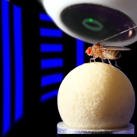
Showing 1-10 of 16 resultsTaste memories allow animals to modulate feeding behavior in accordance with past experience and avoid the consumption of potentially harmful food [1]. We have developed a single-fly taste memory assay to functionally interrogate the neural circuitry encoding taste memories [2]. Here, we screen a collection of Split-GAL4 lines that label small populations of neurons associated with the fly memory center-the mushroom bodies (MBs) [3]. Genetic silencing of PPL1 dopamine neurons disrupts conditioned, but not naive, feeding behavior, suggesting these neurons are selectively involved in the conditioned taste response. We identify two PPL1 subpopulations that innervate the MB α lobe and are essential for aversive taste memory. Thermogenetic activation of these dopamine neurons during training induces memory, indicating these neurons are sufficient for the reinforcing properties of bitter tastant to the MBs. Silencing of either the intrinsic MB neurons or the output neurons from the α lobe disrupts taste conditioning. Thermogenetic manipulation of these output neurons alters naive feeding response, suggesting that dopamine neurons modulate the threshold of response to appetitive tastants. Taken together, these findings detail a neural mechanism underlying the formation of taste memory and provide a functional model for dopamine-dependent plasticity in Drosophila.
Cytotoxic T lymphocytes (CTLs) use polarized secretion to rapidly destroy virally infected and tumor cells. To understand the temporal relationships between key events leading to secretion, we used high-resolution 4D imaging. CTLs approached targets with actin-rich projections at the leading edge, creating an initially actin-enriched contact with rearward-flowing actin. Within 1 min, cortical actin reduced across the synapse, T cell receptors (TCRs) clustered centrally to form the central supramolecular activation cluster (cSMAC), and centrosome polarization began. Granules clustered around the moving centrosome within 2.5 min and reached the synapse after 6 min. TCR-bearing intracellular vesicles were delivered to the cSMAC as the centrosome docked. We found that the centrosome and granules were delivered to an area of membrane with reduced cortical actin density and phospholipid PIP2. These data resolve the temporal order of events during synapse maturation in 4D and reveal a critical role for actin depletion in regulating secretion.
Proper brain function requires the precise localization of proteins and signaling molecules on a nanometer scale. The examination of molecular organization at this scale has been difficult in part because it is beyond the reach of conventional, diffraction-limited light microscopy. The recently developed method of superresolution, localization-based fluorescent microscopy (LBM), such as photoactivated localization microscopy (PALM) and stochastic optical reconstruction microscopy (STORM), has demonstrated a resolving power at a 10 nm scale and is poised to become a vital tool in modern neuroscience research. Indeed, LBM has revealed previously unknown cellular architectures and organizational principles in neurons. Here, we discuss the principles of LBM, its current applications in neuroscience, and the challenges that must be met before its full potential is achieved. We also present the unpublished results of our own experiments to establish a sample preparation procedure for applying LBM to study brain tissue. Synapse, 69:283-294, 2015. © 2015 Wiley Periodicals, Inc.
The neural circuit mechanisms underlying emotion states remain poorly understood. Drosophila offers powerful genetic approaches for dissecting neural circuit function, but whether flies exhibit emotion-like behaviors has not been clear. We recently proposed that model organisms may express internal states displaying “emotion primitives,” which are general characteristics common to different emotions, rather than specific anthropomorphic emotions such as “fear” or “anxiety.” These emotion primitives include scalability, persistence, valence, and generalization to multiple contexts. Here, we have applied this approach to determine whether flies’ defensive responses to moving overhead translational stimuli (“shadows”) are purely reflexive or may express underlying emotion states. We describe a new behavioral assay in which flies confined in an enclosed arena are repeatedly exposed to an overhead translational stimulus. Repetitive stimuli promoted graded (scalable) and persistent increases in locomotor velocity and hopping, and occasional freezing. The stimulus also dispersed feeding flies from a food resource, suggesting both negative valence and context generalization. Strikingly, there was a significant delay before the flies returned to the food following stimulus-induced dispersal, suggestive of a slowly decaying internal defensive state. The length of this delay was increased when more stimuli were delivered for initial dispersal. These responses can be mathematically modeled by assuming an internal state that behaves as a leaky integrator of stimulus exposure. Our results suggest that flies’ responses to repetitive visual threat stimuli express an internal state exhibiting canonical emotion primitives, possibly analogous to fear in mammals. The mechanistic basis of this state can now be investigated in a genetically tractable insect species.
During courtship males attract females with elaborate behaviors. In mice, these displays include ultrasonic vocalizations. Ultrasonic courtship vocalizations were previously attributed to the courting male, despite evidence that both sexes produce virtually indistinguishable vocalizations. Because of this similarity, and the difficulty of assigning vocalizations to individuals, the vocal contribution of each individual during courtship is unknown. To address this question, we developed a microphone array system to localize vocalizations from socially interacting, individual adult mice. With this system, we show that female mice vocally interact with males during courtship. Males and females jointly increased their vocalization rates during chases. Furthermore, a female's participation in these vocal interactions may function as a signal that indicates a state of increased receptivity. Our results reveal a novel form of vocal communication during mouse courtship, and lay the groundwork for a mechanistic dissection of communication during social behavior.
The HMMER website, available at http://www.ebi.ac.uk/Tools/hmmer/, provides access to the protein homology search algorithms found in the HMMER software suite. Since the first release of the website in 2011, the search repertoire has been expanded to include the iterative search algorithm, jackhmmer. The continued growth of the target sequence databases means that traditional tabular representations of significant sequence hits can be overwhelming to the user. Consequently, additional ways of presenting homology search results have been developed, allowing them to be summarised according to taxonomic distribution or domain architecture. The taxonomy and domain architecture representations can be used in combination to filter the results according to the needs of a user. Searches can also be restricted prior to submission using a new taxonomic filter, which not only ensures that the results are specific to the requested taxonomic group, but also improves search performance. The repertoire of profile hidden Markov model libraries, which are used for annotation of query sequences with protein families and domains, has been expanded to include the libraries from CATH-Gene3D, PIRSF, Superfamily and TIGRFAMs. Finally, we discuss the relocation of the HMMER webserver to the European Bioinformatics Institute and the potential impact that this will have.
Observation of molecular processes inside living cells is fundamental to a quantitative understanding of how biological systems function. Specifically, decoding the complex behavior of single molecules enables us to measure kinetics, transport, and self-assembly at this fundamental level that is often veiled in ensemble experiments. In the past decade, rapid developments in fluorescence microscopy, fluorescence correlation spectroscopy, and fluorescent labeling techniques have enabled new experiments to investigate the robustness and stochasticity of diverse molecular mechanisms with high spatiotemporal resolution. This review discusses the concepts and strategies of structural and functional imaging in living cells at the single-molecule level with minimal perturbations to the specimen.
Biological specimens suffer radiation damage when imaged in an electron microscope, ultimately limiting the attainable resolution. At a given resolution, an optimal exposure can be defined that maximizes the signal-to-noise ratio in the image. Using a 2.6 Å resolution single particle cryo-EM reconstruction of rotavirus VP6, determined from movies recorded with a total exposure of 100 electrons/Å(2), we obtained accurate measurements of optimal exposure values over a wide range of resolutions. At low and intermediate resolutions our measured values are considerably higher than obtained previously for crystalline specimens, indicating that both images and movies should be collected with higher exposures than are generally used. We demonstrate a method of using our optimal exposure values to filter movie frames, yielding images with improved contrast that lead to higher resolution reconstructions. This 'high-exposure' technique should benefit cryo-EM work on all types of samples, especially those of relatively low molecular mass.
Many animals navigate using a combination of visual landmarks and path integration. In mammalian brains, head direction cells integrate these two streams of information by representing an animal's heading relative to landmarks, yet maintaining their directional tuning in darkness based on self-motion cues. Here we use two-photon calcium imaging in head-fixed Drosophila melanogaster walking on a ball in a virtual reality arena to demonstrate that landmark-based orientation and angular path integration are combined in the population responses of neurons whose dendrites tile the ellipsoid body, a toroidal structure in the centre of the fly brain. The neural population encodes the fly's azimuth relative to its environment, tracking visual landmarks when available and relying on self-motion cues in darkness. When both visual and self-motion cues are absent, a representation of the animal's orientation is maintained in this network through persistent activity, a potential substrate for short-term memory. Several features of the population dynamics of these neurons and their circular anatomical arrangement are suggestive of ring attractors, network structures that have been proposed to support the function of navigational brain circuits.