Filter
Associated Lab
- Aso Lab (1) Apply Aso Lab filter
- Cui Lab (2) Apply Cui Lab filter
- Harris Lab (1) Apply Harris Lab filter
- Jayaraman Lab (1) Apply Jayaraman Lab filter
- Karpova Lab (1) Apply Karpova Lab filter
- Leonardo Lab (1) Apply Leonardo Lab filter
- Looger Lab (2) Apply Looger Lab filter
- Rubin Lab (3) Apply Rubin Lab filter
- Schreiter Lab (1) Apply Schreiter Lab filter
- Simpson Lab (1) Apply Simpson Lab filter
- Svoboda Lab (1) Apply Svoboda Lab filter
- Tervo Lab (1) Apply Tervo Lab filter
Associated Project Team
Publication Date
- October 25, 2012 (3) Apply October 25, 2012 filter
- October 22, 2012 (1) Apply October 22, 2012 filter
- October 18, 2012 (1) Apply October 18, 2012 filter
- October 12, 2012 (1) Apply October 12, 2012 filter
- October 10, 2012 (1) Apply October 10, 2012 filter
- October 8, 2012 (1) Apply October 8, 2012 filter
- October 5, 2012 (1) Apply October 5, 2012 filter
- October 3, 2012 (1) Apply October 3, 2012 filter
- October 1, 2012 (4) Apply October 1, 2012 filter
- Remove October 2012 filter October 2012
- Remove 2012 filter 2012
14 Janelia Publications
Showing 1-10 of 14 resultsThis paper presents a digital neural/EMG telemetry system small enough and lightweight enough to permit recording from insects in flight. It has a measured flight package mass of only 38 mg. This system includes a single-chip telemetry integrated circuit (IC) employing RF power harvesting for battery-free operation, with communication via modulated backscatter in the UHF (902-928 MHz) band. An on-chip 11-bit ADC digitizes 10 neural channels with a sampling rate of 26.1 kSps and 4 EMG channels at 1.63 kSps, and telemeters this data wirelessly to a base station. The companion base station transceiver includes an RF transmitter of +36 dBm (4 W) output power to wirelessly power the telemetry IC, and a digital receiver with a sensitivity of -70 dBm for 10⁻⁵ BER at 5.0 Mbps to receive the data stream from the telemetry IC. The telemetry chip was fabricated in a commercial 0.35 μ m 4M1P (4 metal, 1 poly) CMOS process. The die measures 2.36 × 1.88 mm, is 250 μm thick, and is wire bonded into a flex circuit assembly measuring 4.6 × 6.8 mm.
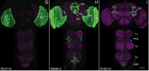
We established a collection of 7,000 transgenic lines of Drosophila melanogaster. Expression of GAL4 in each line is controlled by a different, defined fragment of genomic DNA that serves as a transcriptional enhancer. We used confocal microscopy of dissected nervous systems to determine the expression patterns driven by each fragment in the adult brain and ventral nerve cord. We present image data on 6,650 lines. Using both manual and machine-assisted annotation, we describe the expression patterns in the most useful lines. We illustrate the utility of these data for identifying novel neuronal cell types, revealing brain asymmetry, and describing the nature and extent of neuronal shape stereotypy. The GAL4 lines allow expression of exogenous genes in distinct, small subsets of the adult nervous system. The set of DNA fragments, each driving a documented expression pattern, will facilitate the generation of additional constructs for manipulating neuronal function. synapse was substantially elevated, at the endocytic zone there was no enhanced polymerization activity. We conclude that actin subserves spatially diverse, independently regulated processes throughout spines. Perisynaptic actin forms a uniquely dynamic structure well suited for direct, active regulation of the synapse.
For the overall strategy and methods used to produce the GAL4 lines:
Pfeiffer, B.D., Jenett, A., Hammonds, A.S., Ngo, T.T., Misra, S., Murphy, C., Scully, A., Carlson, J.W., Wan, K.H., Laverty, T.R., Mungall, C., Svirskas, R., Kadonaga, J.T., Doe, C.Q., Eisen, M.B., Celniker, S.E., Rubin, G.M. (2008). Tools for neuroanatomy and neurogenetics in Drosophila. Proc. Natl. Acad. Sci. USA 105, 9715-9720. http://www.pnas.org/content/105/28/9715.full.pdf+html synapse was substantially elevated, at the endocytic zone there was no enhanced polymerization activity. We conclude that actin subserves spatially diverse, independently regulated processes throughout spines. Perisynaptic actin forms a uniquely dynamic structure well suited for direct, active regulation of the synapse.
For data on expression in the embryo:
Manning, L., Purice, M.D., Roberts, J., Pollard, J.L., Bennett, A.L., Kroll, J.R., Dyukareva, A.V., Doan, P.N., Lupton, J.R., Strader, M.E., Tanner, S., Bauer, D., Wilbur, A., Tran, K.D., Laverty, T.R., Pearson, J.C., Crews, S.T., Rubin, G.M., and Doe, C.Q. (2012) Annotated embryonic CNS expression patterns of 5000 GMR GAL4 lines: a resource for manipulating gene expression and analyzing cis-regulatory motifs. Cell Reports (2012) Doi: 10.1016/j.celrep.2012.09.009 http://www.cell.com/cell-reports/fulltext/S2211-1247(12)00290-2 synapse was substantially elevated, at the endocytic zone there was no enhanced polymerization activity. We conclude that actin subserves spatially diverse, independently regulated processes throughout spines. Perisynaptic actin forms a uniquely dynamic structure well suited for direct, active regulation of the synapse.
For data on expression in imaginal discs:
Jory, A., Estella, C., Giorgianni, M.W., Slattery, M., Laverty, T.R., Rubin, G.M., and Mann, R.S. (2012) A survey of 6300 genomic fragments for cis-regulatory activity in the imaginal discs of Drosophila melanogaster. Cell Reports (2012) Doi: 10.1016/j.celrep.2012.09.010 http://www.cell.com/cell-reports/fulltext/S2211-1247(12)00291-4 synapse was substantially elevated, at the endocytic zone there was no enhanced polymerization activity. We conclude that actin subserves spatially diverse, independently regulated processes throughout spines. Perisynaptic actin forms a uniquely dynamic structure well suited for direct, active regulation of the synapse.
For data on expression in the larval nervous system:
Li, H.-H., Kroll, J.R., Lennox, S., Ogundeyi, O., Jeter, J., Depasquale, G., and Truman, J.W. (2013) A GAL4 driver resource for developmental and behavioral studies on the larval CNS of Drosophila. Cell Reports (submitted).
Here, we describe the embryonic central nervous system expression of 5,000 GAL4 lines made using molecularly defined cis-regulatory DNA inserted into a single attP genomic location. We document and annotate the patterns in early embryos when neurogenesis is at its peak, and in older embryos where there is maximal neuronal diversity and the first neural circuits are established. We note expression in other tissues, such as the lateral body wall (muscle, sensory neurons, and trachea) and viscera. Companion papers report on the adult brain and larval imaginal discs, and the integrated data sets are available online (http://www.janelia.org/gal4-gen1). This collection of embryonically expressed GAL4 lines will be valuable for determining neuronal morphology and function. The 1,862 lines expressed in small subsets of neurons (<20/segment) will be especially valuable for characterizing interneuronal diversity and function, because although interneurons comprise the majority of all central nervous system neurons, their gene expression profile and function remain virtually unexplored.
Over 6,000 fragments from the genome of Drosophila melanogaster were analyzed for their ability to drive expression of GAL4 reporter genes in the third-instar larval imaginal discs. About 1,200 reporter genes drove expression in the eye, antenna, leg, wing, haltere, or genital imaginal discs. The patterns ranged from large regions to individual cells. About 75% of the active fragments drove expression in multiple discs; 20% were expressed in ventral, but not dorsal, discs (legs, genital, and antenna), whereas \~{}23% were expressed in dorsal but not ventral discs (wing, haltere, and eye). Several patterns, for example, within the leg chordotonal organ, appeared a surprisingly large number of times. Unbiased searches for DNA sequence motifs suggest candidate transcription factors that may regulate enhancers with shared activities. Together, these expression patterns provide a valuable resource to the community and offer a broad overview of how transcriptional regulatory information is distributed in the Drosophila genome.
Microinfusions of drugs directly into the central nervous system of awake animals represent a widely used means of unravelling brain functions related to behaviour. However, current approaches generally use tethered liquid infusion systems and a syringe pump to deliver drugs into the brain, which often interfere with behaviour. We address this shortfall with a miniaturised electronically-controlled drug delivery system (20 × 17.5 × 5 mm³) designed to be skull-mounted in rats. The device features a micropump connected to two 8-mm-long silicon microprobes with a cross section of 250 × 250 μm² and integrated fluid microchannels. Using an external electronic control unit, the device allows infusion of 16 metered doses (0.25 μL each, 8 per silicon shaft). Each dosage requires 3.375 Ws of electrical power making the device additionally compatible with state-of-the-art wireless headstages. A dosage precision of 0.25 ± 0.01 μL was determined in vitro before in vivo tests were carried out in awake rats. No passive leakage from the loaded devices into the brain could be detected using methylene blue dye. Finally, the device was used to investigate the effects of the NMDA-receptor antagonist 3-((R)-2-Carboxypiperazin-4-yl)-propyl-1-phosphonic acid, (R)-CPP, administered directly into the prefrontal cortex of rats during performance on a task to assess visual attention and impulsivity. In agreement with previous findings using conventional tethered infusion systems, acute (R)-CPP administration produced a marked increase in impulsivity.
Optical microscopy has so far been restricted to superficial layers, leaving many important biological questions unanswered. Random scattering causes the ballistic focus, which is conventionally used for image formation, to decay exponentially with depth. Optical imaging beyond the ballistic regime has been demonstrated by hybrid techniques that combine light with the deeper penetration capability of sound waves. Deep inside highly scattering media, the sound focus dimensions restrict the imaging resolutions. Here we show that by iteratively focusing light into an ultrasound focus via phase conjugation, we can fundamentally overcome this resolution barrier in deep tissues and at the same time increase the focus to background ratio. We demonstrate fluorescence microscopy beyond the ballistic regime of light with a threefold improved resolution and a fivefold increase in contrast. This development opens up practical high resolution fluorescence imaging in deep tissues.
Many tools are available to analyse genomes but are often challenging to use in a cell type-specific context. We have developed a method similar to the isolation of nuclei tagged in a specific cell type (INTACT) technique [Deal,R.B. and Henikoff,S. (2010) A simple method for gene expression and chromatin profiling of individual cell types within a tissue. Dev. Cell, 18, 1030-1040; Steiner,F.A., Talbert,P.B., Kasinathan,S., Deal,R.B. and Henikoff,S. (2012) Cell-type-specific nuclei purification from whole animals for genome-wide expression and chromatin profiling. Genome Res., doi:10.1101/gr.131748.111], first developed in plants, for use in Drosophila neurons. We profile gene expression and histone modifications in Kenyon cells and octopaminergic neurons in the adult brain. In addition to recovering known gene expression differences, we also observe significant cell type-specific chromatin modifications. In particular, a small subset of differentially expressed genes exhibits a striking anti-correlation between repressive and activating histone modifications. These genes are enriched for transcription factors, recovering those known to regulate mushroom body identity and predicting analogous regulators of octopaminergic neurons. Our results suggest that applying INTACT to specific neuronal populations can illuminate the transcriptional regulatory networks that underlie neuronal cell identity.
The GABA transporters (GAT1, GAT2, GAT3, and BGT1) have mostly been discussed in relation to their potential roles in controlling the action of transmitter GABA in the nervous system. We have generated the first mice lacking the GAT2 (slc6a13) gene. Deletion of GAT2 (both mRNA and protein) neither affected growth, fertility, nor life span under nonchallenging rearing conditions. Immunocytochemistry showed that the GAT2 protein was predominantly expressed in the plasma membranes of periportal hepatocytes and in the basolateral membranes of proximal tubules in the renal cortex. This was validated by processing tissue from wild-type and knockout mice in parallel. Deletion of GAT2 reduced liver taurine levels by 50%, without affecting the expression of the taurine transporter TAUT. These results suggest an important role for GAT2 in taurine uptake from portal blood into liver. In support of this notion, GAT2-transfected HEK293 cells transported [(3)H]taurine. Furthermore, most of the uptake of [(3)H]GABA by cultured rat hepatocytes was due to GAT2, and this uptake was inhibited by taurine. GAT2 was not detected in brain parenchyma proper, excluding a role in GABA inactivation. It was, however, expressed in the leptomeninges and in a subpopulation of brain blood vessels. Deletion of GAT2 increased brain taurine levels by 20%, suggesting a taurine-exporting role for GAT2 in the brain.
The olfactory system encodes information about molecules by spatiotemporal patterns of activity across distributed populations of neurons and extracts information from these patterns to control specific behaviors. Recent studies used in vivo recordings, optogenetics, and other methods to analyze the mechanisms by which odor information is encoded and processed in the olfactory system, the functional connectivity within and between olfactory brain areas, and the impact of spatiotemporal patterning of neuronal activity on higher-order neurons and behavioral outputs. The results give rise to a faceted picture of olfactory processing and provide insights into fundamental mechanisms underlying neuronal computations. This review focuses on some of this work presented in a Mini-Symposium at the Annual Meeting of the Society for Neuroscience in 2012.
The ability to chronically monitor neuronal activity in the living brain is essential for understanding the organization and function of the nervous system. The genetically encoded green fluorescent protein based calcium sensor GCaMP provides a powerful tool for detecting calcium transients in neuronal somata, processes, and synapses that are triggered by neuronal activities. Here we report the generation and characterization of transgenic mice that express improved GCaMPs in various neuronal subpopulations under the control of the Thy1 promoter. In vitro and in vivo studies show that calcium transients induced by spontaneous and stimulus-evoked neuronal activities can be readily detected at the level of individual cells and synapses in acute brain slices, as well as in awake behaving animals. These GCaMP transgenic mice allow investigation of activity patterns in defined neuronal populations in the living brain, and will greatly facilitate dissecting complex structural and functional relationships of neural networks.