Filter
Associated Lab
- Betzig Lab (8) Apply Betzig Lab filter
- Eddy/Rivas Lab (2) Apply Eddy/Rivas Lab filter
- Fetter Lab (1) Apply Fetter Lab filter
- Hess Lab (2) Apply Hess Lab filter
- Ji Lab (4) Apply Ji Lab filter
- Looger Lab (3) Apply Looger Lab filter
- Magee Lab (4) Apply Magee Lab filter
- Riddiford Lab (1) Apply Riddiford Lab filter
- Rubin Lab (2) Apply Rubin Lab filter
- Schreiter Lab (1) Apply Schreiter Lab filter
- Shroff Lab (6) Apply Shroff Lab filter
- Spruston Lab (3) Apply Spruston Lab filter
- Sternson Lab (1) Apply Sternson Lab filter
- Svoboda Lab (6) Apply Svoboda Lab filter
- Truman Lab (1) Apply Truman Lab filter
Publication Date
- December 2008 (8) Apply December 2008 filter
- November 2008 (1) Apply November 2008 filter
- October 2008 (1) Apply October 2008 filter
- September 2008 (2) Apply September 2008 filter
- August 2008 (3) Apply August 2008 filter
- July 2008 (4) Apply July 2008 filter
- June 2008 (1) Apply June 2008 filter
- May 2008 (3) Apply May 2008 filter
- April 2008 (1) Apply April 2008 filter
- March 2008 (5) Apply March 2008 filter
- February 2008 (5) Apply February 2008 filter
- January 2008 (6) Apply January 2008 filter
- Remove 2008 filter 2008
40 Janelia Publications
Showing 31-40 of 40 resultsThe chemical senses, smell and taste, are the most poorly understood sensory modalities. In recent years, however, the field of chemosensation has benefited from new methods and technical innovations that have accelerated the rate of scientific progress. For example, enormous advances have been made in identifying olfactory and gustatory receptor genes and mapping their expression patterns. Genetic tools now permit us to monitor and control neural activity in vivo with unprecedented precision. New imaging techniques allow us to watch neural activity patterns unfold in real time. Finally, improved hardware and software enable multineuron electrophysiological recordings on an expanded scale. These innovations have enabled some fresh approaches to classic problems in chemosensation.
Genetically encoded calcium indicators (GECIs), based on recombinant fluorescent proteins, have been engineered to observe calcium transients in living cells and organisms. Through observation of calcium, these indicators also report neural activity. We review progress in GECI construction and application, particularly toward in vivo monitoring of sparse action potentials (APs). We summarize the extrinsic and intrinsic factors that influence GECI performance. A simple model of GECI response to AP firing demonstrates the relative significance of these factors. We recommend a standardized protocol for evaluating GECIs in a physiologically relevant context. A potential method of simultaneous optical control and recording of neuronal circuits is presented.
Electrical microstimulation can establish causal links between the activity of groups of neurons and perceptual and cognitive functions. However, the number and identities of neurons microstimulated, as well as the number of action potentials evoked, are difficult to ascertain. To address these issues we introduced the light-gated algal channel channelrhodopsin-2 (ChR2) specifically into a small fraction of layer 2/3 neurons of the mouse primary somatosensory cortex. ChR2 photostimulation in vivo reliably generated stimulus-locked action potentials at frequencies up to 50 Hz. Here we show that naive mice readily learned to detect brief trains of action potentials (five light pulses, 1 ms, 20 Hz). After training, mice could detect a photostimulus firing a single action potential in approximately 300 neurons. Even fewer neurons (approximately 60) were required for longer stimuli (five action potentials, 250 ms). Our results show that perceptual decisions and learning can be driven by extremely brief epochs of cortical activity in a sparse subset of supragranular cortical pyramidal neurons.
MOTIVATION: Caenorhabditis elegans, a roundworm found in soil, is a widely studied model organism with about 1000 cells in the adult. Producing high-resolution fluorescence images of C.elegans to reveal biological insights is becoming routine, motivating the development of advanced computational tools for analyzing the resulting image stacks. For example, worm bodies usually curve significantly in images. Thus one must ’straighten’ the worms if they are to be compared under a canonical coordinate system. RESULTS: We develop a worm straightening algorithm (WSA) that restacks cutting planes orthogonal to a ’backbone’ that models the anterior-posterior axis of the worm. We formulate the backbone as a parametric cubic spline defined by a series of control points. We develop two methods for automatically determining the locations of the control points. Our experimental methods show that our approaches effectively straighten both 2D and 3D worm images.
Pfam is a comprehensive collection of protein domains and families, represented as multiple sequence alignments and as profile hidden Markov models. The current release of Pfam (22.0) contains 9318 protein families. Pfam is now based not only on the UniProtKB sequence database, but also on NCBI GenPept and on sequences from selected metagenomics projects. Pfam is available on the web from the consortium members using a new, consistent and improved website design in the UK (http://pfam.sanger.ac.uk/), the USA (http://pfam.janelia.org/) and Sweden (http://pfam.sbc.su.se/), as well as from mirror sites in France (http://pfam.jouy.inra.fr/) and South Korea (http://pfam.ccbb.re.kr/).
In neurons, individual dendritic spines isolate N-methyl-d-aspartate (NMDA) receptor-mediated calcium ion (Ca2+) accumulations from the dendrite and other spines. However, the extent to which spines compartmentalize signaling events downstream of Ca2+ influx is not known. We combined two-photon fluorescence lifetime imaging with two-photon glutamate uncaging to image the activity of the small guanosine triphosphatase Ras after NMDA receptor activation at individual spines. Induction of long-term potentiation (LTP) triggered robust Ca2+-dependent Ras activation in single spines that decayed in approximately 5 minutes. Ras activity spread over approximately 10 micrometers of dendrite and invaded neighboring spines by diffusion. The spread of Ras-dependent signaling was necessary for the local regulation of the threshold for LTP induction. Thus, Ca2+-dependent synaptic signals can spread to couple multiple synapses on short stretches of dendrite.
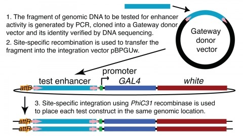
We demonstrate the feasibility of generating thousands of transgenic Drosophila melanogaster lines in which the expression of an exogenous gene is reproducibly directed to distinct small subsets of cells in the adult brain. We expect the expression patterns produced by the collection of 5,000 lines that we are currently generating to encompass all neurons in the brain in a variety of intersecting patterns. Overlapping 3-kb DNA fragments from the flanking noncoding and intronic regions of genes thought to have patterned expression in the adult brain were inserted into a defined genomic location by site-specific recombination. These fragments were then assayed for their ability to function as transcriptional enhancers in conjunction with a synthetic core promoter designed to work with a wide variety of enhancer types. An analysis of 44 fragments from four genes found that >80% drive expression patterns in the brain; the observed patterns were, on average, comprised of <100 cells. Our results suggest that the D. melanogaster genome contains >50,000 enhancers and that multiple enhancers drive distinct subsets of expression of a gene in each tissue and developmental stage. We expect that these lines will be valuable tools for neuroanatomy as well as for the elucidation of neuronal circuits and information flow in the fly brain.
In practice, understanding the spatial relationships between the surfaces of an object, can significantly improve the performance of object recognition systems. In this paper we propose a novel framework to recognize objects in pictures taken from arbitrary viewpoints. The idea is to maintain the frontal views of the major faces of objects in a global flat map. Then an unfolding warping technique is used to change the pose of the query object in the test view so that all visible surfaces of the object can be observed from a frontal viewpoint, improving the handling of serious occlusions and large viewpoint changes. We demonstrate the effectiveness of our approach through analysis of recognition trials of complex objects with comparison to popular methods.
The conditional expression of hairpin constructs in Drosophila melanogaster has emerged in recent years as a method of choice in functional genomic studies. To date, upstream activating site-driven RNA interference constructs have been inserted into the genome randomly using P-element-mediated transformation, which can result in false negatives due to variable expression. To avoid this problem, we have developed a transgenic RNA interference vector based on the phiC31 site-specific integration method.