Filter
Associated Lab
- Aso Lab (1) Apply Aso Lab filter
- Baker Lab (3) Apply Baker Lab filter
- Betzig Lab (7) Apply Betzig Lab filter
- Bock Lab (1) Apply Bock Lab filter
- Cui Lab (3) Apply Cui Lab filter
- Dickson Lab (1) Apply Dickson Lab filter
- Dudman Lab (1) Apply Dudman Lab filter
- Eddy/Rivas Lab (5) Apply Eddy/Rivas Lab filter
- Fetter Lab (2) Apply Fetter Lab filter
- Gonen Lab (1) Apply Gonen Lab filter
- Hess Lab (2) Apply Hess Lab filter
- Jayaraman Lab (2) Apply Jayaraman Lab filter
- Ji Lab (2) Apply Ji Lab filter
- Keller Lab (2) Apply Keller Lab filter
- Lavis Lab (5) Apply Lavis Lab filter
- Lee (Albert) Lab (1) Apply Lee (Albert) Lab filter
- Leonardo Lab (2) Apply Leonardo Lab filter
- Looger Lab (7) Apply Looger Lab filter
- Magee Lab (1) Apply Magee Lab filter
- Menon Lab (3) Apply Menon Lab filter
- Murphy Lab (1) Apply Murphy Lab filter
- Reiser Lab (2) Apply Reiser Lab filter
- Riddiford Lab (1) Apply Riddiford Lab filter
- Rubin Lab (3) Apply Rubin Lab filter
- Scheffer Lab (2) Apply Scheffer Lab filter
- Schreiter Lab (2) Apply Schreiter Lab filter
- Simpson Lab (3) Apply Simpson Lab filter
- Singer Lab (1) Apply Singer Lab filter
- Sternson Lab (6) Apply Sternson Lab filter
- Svoboda Lab (7) Apply Svoboda Lab filter
- Tjian Lab (2) Apply Tjian Lab filter
- Truman Lab (1) Apply Truman Lab filter
- Zlatic Lab (1) Apply Zlatic Lab filter
- Zuker Lab (2) Apply Zuker Lab filter
Associated Project Team
Publication Date
- December 2011 (10) Apply December 2011 filter
- November 2011 (8) Apply November 2011 filter
- October 2011 (8) Apply October 2011 filter
- September 2011 (8) Apply September 2011 filter
- August 2011 (9) Apply August 2011 filter
- July 2011 (5) Apply July 2011 filter
- June 2011 (10) Apply June 2011 filter
- May 2011 (6) Apply May 2011 filter
- April 2011 (5) Apply April 2011 filter
- March 2011 (6) Apply March 2011 filter
- February 2011 (8) Apply February 2011 filter
- January 2011 (15) Apply January 2011 filter
- Remove 2011 filter 2011
98 Janelia Publications
Showing 91-98 of 98 resultsThermosensation is an indispensable sensory modality. Here, we study temperature coding in Drosophila, and show that temperature is represented by a spatial map of activity in the brain. First, we identify TRP channels that function in the fly antenna to mediate the detection of cold stimuli. Next, we identify the hot-sensing neurons and show that hot and cold antennal receptors project onto distinct, but adjacent glomeruli in the Proximal-Antennal-Protocerebrum (PAP) forming a thermotopic map in the brain. We use two-photon imaging to reveal the functional segregation of hot and cold responses in the PAP, and show that silencing the hot- or cold-sensing neurons produces animals with distinct and discrete deficits in their behavioral responses to thermal stimuli. Together, these results demonstrate that dedicated populations of cells orchestrate behavioral responses to different temperature stimuli, and reveal a labeled-line logic for the coding of temperature information in the brain.
The innate sexual behaviors of Drosophila melanogaster males are an attractive system for elucidating how complex behavior patterns are generated. The potential for male sexual behavior in D. melanogaster is specified by the fruitless (fru) and doublesex (dsx) sex regulatory genes. We used the temperature-sensitive activator dTRPA1 to probe the roles of fru(M)- and dsx-expressing neurons in male courtship behaviors. Almost all steps of courtship, from courtship song to ejaculation, can be induced at very high levels through activation of either all fru(M) or all dsx neurons in solitary males. Detailed characterizations reveal different roles for fru(M) and dsx in male courtship. Surprisingly, the system for mate discrimination still works well when all dsx neurons are activated, but is impaired when all fru(M) neurons are activated. Most strikingly, we provide evidence for a fru(M)-independent courtship pathway that is primarily vision dependent.
Multiphoton imaging (MPI) is widely used for recording activity simultaneously from many neurons in superficial cortical layers in vivo. We combined regenerative amplification multiphoton microscopy (RAMM) with genetically encoded calcium indicators to extend MPI of neuronal population activity into layer 5 (L5) of adult mouse somatosensory cortex. We found that this approach could be used to record and quantify spontaneous and sensory-evoked activity in populations of L5 neuronal somata located as much as 800 μm below the pia. In addition, we found that RAMM could be used to simultaneously image activity from large (80) populations of apical dendrites and follow these dendrites down to their somata of origin.
The ability of insects to learn and navigate to specific locations in the environment has fascinated naturalists for decades. The impressive navigational abilities of ants, bees, wasps and other insects demonstrate that insects are capable of visual place learning, but little is known about the underlying neural circuits that mediate these behaviours. Drosophila melanogaster (common fruit fly) is a powerful model organism for dissecting the neural circuitry underlying complex behaviours, from sensory perception to learning and memory. Drosophila can identify and remember visual features such as size, colour and contour orientation. However, the extent to which they use vision to recall specific locations remains unclear. Here we describe a visual place learning platform and demonstrate that Drosophila are capable of forming and retaining visual place memories to guide selective navigation. By targeted genetic silencing of small subsets of cells in the Drosophila brain, we show that neurons in the ellipsoid body, but not in the mushroom bodies, are necessary for visual place learning. Together, these studies reveal distinct neuroanatomical substrates for spatial versus non-spatial learning, and establish Drosophila as a powerful model for the study of spatial memories.
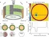
We have developed miniature telemetry systems that capture neural, EMG, and acceleration signals from a freely moving insect or other small animal and transmit the data wirelessly to a remote digital receiver. The systems are based on custom low-power integrated circuits (ICs) that amplify, filter, and digitize four biopotential signals using low-noise circuits. One of the chips also digitizes three acceleration signals from an off-chip microelectromechanical-system accelerometer. All information is transmitted over a wireless ~ 900-MHz telemetry link. The first unit, using a custom chip fabricated in a 0.6- μm BiCMOS process, weighs 0.79 g and runs for two hours on two small batteries. We have used this system to monitor neural and EMG signals in jumping and flying locusts as well as transdermal potentials in weakly swimming electric fish. The second unit, using a custom chip fabricated in a 0.35-μ m complementary metal-oxide semiconductor CMOS process, weighs 0.17 g and runs for five hours on a single 1.5-V battery. This system has been used to monitor neural potentials in untethered perching dragonflies.
Wiring economy has successfully explained the individual placement of neurons in simple nervous systems like that of Caenorhabditis elegans [1-3] and the locations of coarser structures like cortical areas in complex vertebrate brains [4]. However, it remains unclear whether wiring economy can explain the placement of individual neurons in brains larger than that of C. elegans. Indeed, given the greater number of neuronal interconnections in larger brains, simply minimizing the length of connections results in unrealistic configurations, with multiple neurons occupying the same position in space. Avoiding such configurations, or volume exclusion, repels neurons from each other, thus counteracting wiring economy. Here we test whether wiring economy together with volume exclusion can explain the placement of neurons in a module of the Drosophila melanogaster brain known as lamina cartridge [5-13]. We used newly developed techniques for semiautomated reconstruction from serial electron microscopy (EM) [14] to obtain the shapes of neurons, the location of synapses, and the resultant synaptic connectivity. We show that wiring length minimization and volume exclusion together can explain the structure of the lamina microcircuit. Therefore, even in brains larger than that of C. elegans, at least for some circuits, optimization can play an important role in individual neuron placement.