Filter
Associated Lab
- Ahrens Lab (3) Apply Ahrens Lab filter
- Aso Lab (6) Apply Aso Lab filter
- Baker Lab (2) Apply Baker Lab filter
- Betzig Lab (11) Apply Betzig Lab filter
- Branson Lab (6) Apply Branson Lab filter
- Cardona Lab (5) Apply Cardona Lab filter
- Chklovskii Lab (2) Apply Chklovskii Lab filter
- Cui Lab (5) Apply Cui Lab filter
- Dickson Lab (1) Apply Dickson Lab filter
- Druckmann Lab (3) Apply Druckmann Lab filter
- Dudman Lab (3) Apply Dudman Lab filter
- Eddy/Rivas Lab (4) Apply Eddy/Rivas Lab filter
- Egnor Lab (1) Apply Egnor Lab filter
- Fetter Lab (5) Apply Fetter Lab filter
- Freeman Lab (7) Apply Freeman Lab filter
- Funke Lab (1) Apply Funke Lab filter
- Gonen Lab (5) Apply Gonen Lab filter
- Grigorieff Lab (7) Apply Grigorieff Lab filter
- Harris Lab (7) Apply Harris Lab filter
- Heberlein Lab (1) Apply Heberlein Lab filter
- Hess Lab (7) Apply Hess Lab filter
- Jayaraman Lab (4) Apply Jayaraman Lab filter
- Ji Lab (4) Apply Ji Lab filter
- Karpova Lab (1) Apply Karpova Lab filter
- Keleman Lab (2) Apply Keleman Lab filter
- Keller Lab (7) Apply Keller Lab filter
- Lavis Lab (5) Apply Lavis Lab filter
- Leonardo Lab (2) Apply Leonardo Lab filter
- Liu (Zhe) Lab (4) Apply Liu (Zhe) Lab filter
- Looger Lab (9) Apply Looger Lab filter
- Magee Lab (5) Apply Magee Lab filter
- Murphy Lab (1) Apply Murphy Lab filter
- Pastalkova Lab (3) Apply Pastalkova Lab filter
- Reiser Lab (2) Apply Reiser Lab filter
- Romani Lab (2) Apply Romani Lab filter
- Rubin Lab (16) Apply Rubin Lab filter
- Saalfeld Lab (3) Apply Saalfeld Lab filter
- Scheffer Lab (2) Apply Scheffer Lab filter
- Schreiter Lab (4) Apply Schreiter Lab filter
- Shroff Lab (1) Apply Shroff Lab filter
- Simpson Lab (4) Apply Simpson Lab filter
- Singer Lab (10) Apply Singer Lab filter
- Spruston Lab (7) Apply Spruston Lab filter
- Stern Lab (4) Apply Stern Lab filter
- Sternson Lab (7) Apply Sternson Lab filter
- Svoboda Lab (9) Apply Svoboda Lab filter
- Tjian Lab (6) Apply Tjian Lab filter
- Truman Lab (6) Apply Truman Lab filter
- Turaga Lab (1) Apply Turaga Lab filter
- Turner Lab (2) Apply Turner Lab filter
- Wu Lab (1) Apply Wu Lab filter
- Zlatic Lab (4) Apply Zlatic Lab filter
- Zuker Lab (2) Apply Zuker Lab filter
Associated Project Team
Associated Support Team
- Electron Microscopy (1) Apply Electron Microscopy filter
- Gene Targeting and Transgenics (3) Apply Gene Targeting and Transgenics filter
- Invertebrate Shared Resource (1) Apply Invertebrate Shared Resource filter
- Janelia Experimental Technology (2) Apply Janelia Experimental Technology filter
- Management Team (1) Apply Management Team filter
- Molecular Genomics (1) Apply Molecular Genomics filter
- Primary & iPS Cell Culture (2) Apply Primary & iPS Cell Culture filter
- Project Technical Resources (1) Apply Project Technical Resources filter
- Scientific Computing Software (11) Apply Scientific Computing Software filter
Publication Date
- December 2015 (15) Apply December 2015 filter
- November 2015 (22) Apply November 2015 filter
- October 2015 (16) Apply October 2015 filter
- September 2015 (16) Apply September 2015 filter
- August 2015 (17) Apply August 2015 filter
- July 2015 (18) Apply July 2015 filter
- June 2015 (16) Apply June 2015 filter
- May 2015 (16) Apply May 2015 filter
- April 2015 (18) Apply April 2015 filter
- March 2015 (16) Apply March 2015 filter
- February 2015 (15) Apply February 2015 filter
- January 2015 (10) Apply January 2015 filter
- Remove 2015 filter 2015
195 Janelia Publications
Showing 71-80 of 195 resultsThe RNA-guided CRISPR-associated protein Cas9 is used for genome editing, transcriptional modulation, and live-cell imaging. Cas9-guide RNA complexes recognize and cleave double-stranded DNA sequences on the basis of 20-nucleotide RNA-DNA complementarity, but the mechanism of target searching in mammalian cells is unknown. Here, we use single-particle tracking to visualize diffusion and chromatin binding of Cas9 in living cells. We show that three-dimensional diffusion dominates Cas9 searching in vivo, and off-target binding events are, on average, short-lived (<1 second). Searching is dependent on the local chromatin environment, with less sampling and slower movement within heterochromatin. These results reveal how the bacterial Cas9 protein interrogates mammalian genomes and navigates eukaryotic chromatin structure.
Cortical spreading depression is a slowly propagating wave of near-complete depolarization of brain cells followed by temporary suppression of neuronal activity. Accumulating evidence indicates that cortical spreading depression underlies the migraine aura and that similar waves promote tissue damage in stroke, trauma, and hemorrhage. Cortical spreading depression is characterized by neuronal swelling, profound elevation of extracellular potassium and glutamate, multiphasic blood flow changes, and drop in tissue oxygen tension. The slow speed of the cortical spreading depression wave implies that it is mediated by diffusion of a chemical substance, yet the identity of this substance and the pathway it follows are unknown. Intercellular spread between gap junction-coupled neurons or glial cells and interstitial diffusion of K(+) or glutamate have been proposed. Here we use extracellular direct current potential recordings, K(+)-sensitive microelectrodes, and 2-photon imaging with ultrasensitive Ca(2+) and glutamate fluorescent probes to elucidate the spatiotemporal dynamics of ionic shifts associated with the propagation of cortical spreading depression in the visual cortex of adult living mice. Our data argue against intercellular spread of Ca(2+) carrying the cortical spreading depression wavefront and are in favor of interstitial K(+) diffusion, rather than glutamate diffusion, as the leading event in cortical spreading depression.
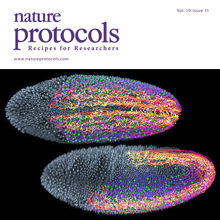
Light-sheet microscopy is a powerful method for imaging the development and function of complex biological systems at high spatiotemporal resolution and over long time scales. Such experiments typically generate terabytes of multidimensional image data, and thus they demand efficient computational solutions for data management, processing and analysis. We present protocols and software to tackle these steps, focusing on the imaging-based study of animal development. Our protocols facilitate (i) high-speed lossless data compression and content-based multiview image fusion optimized for multicore CPU architectures, reducing image data size 30–500-fold; (ii) automated large-scale cell tracking and segmentation; and (iii) visualization, editing and annotation of multiterabyte image data and cell-lineage reconstructions with tens of millions of data points. These software modules are open source. They provide high data throughput using a single computer workstation and are readily applicable to a wide spectrum of biological model systems.
Emotional processes are central to behavior, yet their deeply subjective nature has been a challenge for neuroscientific study as well as for psychiatric diagnosis. Here we explore the relationships between subjective feelings and their underlying brain circuits from a computational perspective. We apply recent insights from systems neuroscience-approaching subjective behavior as the result of mental computations instantiated in the brain-to the study of emotions. We develop the hypothesis that emotions are the product of neural computations whose motor role is to reallocate bodily resources mostly gated by smooth muscles. This "emotor" control system is analagous to the more familiar motor control computations that coordinate skeletal muscle movements. To illustrate this framework, we review recent research on "confidence." Although familiar as a feeling, confidence is also an objective statistical quantity: an estimate of the probability that a hypothesis is correct. This model-based approach helped reveal the neural basis of decision confidence in mammals and provides a bridge to the subjective feeling of confidence in humans. These results have important implications for psychiatry, since disorders of confidence computations appear to contribute to a number of psychopathologies. More broadly, this computational approach to emotions resonates with the emerging view that psychiatric nosology may be best parameterized in terms of disorders of the cognitive computations underlying complex behavior.
There is considerable potential for X-ray free electron lasers (XFELs) to enable determination of macromolecular crystal structures that are difficult to solve using current synchrotron sources. Prior XFEL studies often involved the collection of thousands to millions of diffraction images, in part due to limitations of data processing methods. We implemented a data processing system based on classical post-refinement techniques, adapted to specific properties of XFEL diffraction data. When applied to XFEL data from three different proteins collected using various sample delivery systems and XFEL beam parameters, our method improved the quality of the diffraction data as well as the resulting refined atomic models and electron density maps. Moreover, the number of observations for a reflection necessary to assemble an accurate data set could be reduced to a few observations. These developments will help expand the applicability of XFEL crystallography to challenging biological systems, including cases where sample is limited.
Neuronal diversity is essential for mammalian brain function but poses a challenge to molecular profiling. To address the need for tools that facilitate cell-type-specific epigenomic studies, we developed the first affinity purification approach to isolate nuclei from genetically defined cell types in a mammal. We combine this technique with next-generation sequencing to show that three subtypes of neocortical neurons have highly distinctive epigenomic landscapes. Over 200,000 regions differ in chromatin accessibility and DNA methylation signatures characteristic of gene regulatory regions. By footprinting and motif analyses, these regions are predicted to bind distinct cohorts of neuron subtype-specific transcription factors. Neuronal epigenomes reflect both past and present gene expression, with DNA hyper-methylation at developmentally critical genes appearing as a novel epigenomic signature in mature neurons. Taken together, our findings link the functional and transcriptional complexity of neurons to their underlying epigenomic diversity.
The K2 Summit camera was initially the only commercially available direct electron detection camera that was optimized for high-speed counting of primary electrons and was also the only one that implemented centroiding so that the resolution of the camera can be extended beyond the Nyquist limit set by the physical pixel size. In this study, we used well-characterized two-dimensional crystals of the membrane protein aquaporin-0 to characterize the performance of the camera below and beyond the physical Nyquist limit and to measure the influence of electron dose rate on image amplitudes and phases.
Bilaterally symmetric motor patterns-those in which left-right pairs of muscles contract synchronously and with equal amplitude (such as breathing, smiling, whisking, and locomotion)-are widespread throughout the animal kingdom. Yet, surprisingly little is known about the underlying neural circuits. We performed a thermogenetic screen to identify neurons required for bilaterally symmetric locomotion in Drosophila larvae and identified the evolutionarily conserved Even-skipped(+) interneurons (Eve/Evx). Activation or ablation of Eve(+) interneurons disrupted bilaterally symmetric muscle contraction amplitude, without affecting the timing of motor output. Eve(+) interneurons are not rhythmically active and thus function independently of the locomotor CPG. GCaMP6 calcium imaging of Eve(+) interneurons in freely moving larvae showed left-right asymmetric activation that correlated with larval behavior. TEM reconstruction of Eve(+) interneuron inputs and outputs showed that the Eve(+) interneurons are at the core of a sensorimotor circuit capable of detecting and modifying body wall muscle contraction.
Novel body structures are often generated by the redeployment of ancestral components of the genome. In this issue of Developmental Cell, Glassford et al. (2015) present a thorough analysis of the co-option of a gene regulatory network in the origin of an evolutionary novelty.