Filter
Associated Lab
- Ahrens Lab (1) Apply Ahrens Lab filter
- Aso Lab (4) Apply Aso Lab filter
- Remove Branson Lab filter Branson Lab
- Card Lab (3) Apply Card Lab filter
- Cardona Lab (1) Apply Cardona Lab filter
- Dickson Lab (1) Apply Dickson Lab filter
- Dudman Lab (1) Apply Dudman Lab filter
- Fetter Lab (1) Apply Fetter Lab filter
- Freeman Lab (2) Apply Freeman Lab filter
- Harris Lab (1) Apply Harris Lab filter
- Heberlein Lab (1) Apply Heberlein Lab filter
- Jayaraman Lab (1) Apply Jayaraman Lab filter
- Karpova Lab (1) Apply Karpova Lab filter
- Keller Lab (3) Apply Keller Lab filter
- Otopalik Lab (1) Apply Otopalik Lab filter
- Pachitariu Lab (1) Apply Pachitariu Lab filter
- Reiser Lab (4) Apply Reiser Lab filter
- Rubin Lab (8) Apply Rubin Lab filter
- Simpson Lab (1) Apply Simpson Lab filter
- Svoboda Lab (1) Apply Svoboda Lab filter
- Tervo Lab (1) Apply Tervo Lab filter
- Truman Lab (1) Apply Truman Lab filter
- Turaga Lab (5) Apply Turaga Lab filter
- Zlatic Lab (1) Apply Zlatic Lab filter
Associated Project Team
Associated Support Team
- Project Pipeline Support (1) Apply Project Pipeline Support filter
- Anatomy and Histology (1) Apply Anatomy and Histology filter
- Electron Microscopy (1) Apply Electron Microscopy filter
- Invertebrate Shared Resource (2) Apply Invertebrate Shared Resource filter
- Janelia Experimental Technology (1) Apply Janelia Experimental Technology filter
- Project Technical Resources (6) Apply Project Technical Resources filter
- Quantitative Genomics (1) Apply Quantitative Genomics filter
- Scientific Computing Software (6) Apply Scientific Computing Software filter
- Scientific Computing Systems (1) Apply Scientific Computing Systems filter
Publication Date
- 2025 (4) Apply 2025 filter
- 2024 (4) Apply 2024 filter
- 2023 (2) Apply 2023 filter
- 2021 (2) Apply 2021 filter
- 2020 (3) Apply 2020 filter
- 2019 (3) Apply 2019 filter
- 2018 (5) Apply 2018 filter
- 2017 (7) Apply 2017 filter
- 2016 (5) Apply 2016 filter
- 2015 (6) Apply 2015 filter
- 2014 (6) Apply 2014 filter
- 2012 (3) Apply 2012 filter
50 Janelia Publications
Showing 31-40 of 50 resultsRecent developments in machine vision methods for automatic, quantitative analysis of social behavior have immensely improved both the scale and level of resolution with which we can dissect interactions between members of the same species. In this paper, we review these methods, with a particular focus on how biologists can apply them to their own work. We discuss several components of machine vision-based analyses: methods to record high-quality video for automated analyses, video-based tracking algorithms for estimating the positions of interacting animals, and machine learning methods for recognizing patterns of interactions. These methods are extremely general in their applicability, and we review a subset of successful applications of them to biological questions in several model systems with very different types of social behaviors.

Assigning behavioral functions to neural structures has long been a central goal in neuroscience and is a necessary first step toward a circuit-level understanding of how the brain generates behavior. Here, we map the neural substrates of locomotion and social behaviors for Drosophila melanogaster using automated machine-vision and machine-learning techniques. From videos of 400,000 flies, we quantified the behavioral effects of activating 2,204 genetically targeted populations of neurons. We combined a novel quantification of anatomy with our behavioral analysis to create brain-behavior correlation maps, which are shared as browsable web pages and interactive software. Based on these maps, we generated hypotheses of regions of the brain causally related to sensory processing, locomotor control, courtship, aggression, and sleep. Our maps directly specify genetic tools to target these regions, which we used to identify a small population of neurons with a role in the control of walking. •We developed machine-vision methods to broadly and precisely quantify fly behavior•We measured effects of activating 2,204 genetically targeted neuronal populations•We created whole-brain maps of neural substrates of locomotor and social behaviors•We created resources for exploring our results and enabling further investigation Machine-vision analyses of large behavior and neuroanatomy data reveal whole-brain maps of regions associated with numerous complex behaviors.
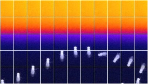
Insects, like most animals, tend to steer away from imminent threats [1-7]. Drosophila melanogaster, for example, generally initiate an escape take-off in response to a looming visual stimulus, mimicking a potential predator [8]. The escape response to a visual threat is, however, flexible [9-12] and can alternatively consist of walking backward away from the perceived threat [11], which may be a more effective response to ambush predators such as nymphal praying mantids [7]. Flexibility in escape behavior may also add an element of unpredictability that makes it difficult for predators to anticipate or learn the prey's likely response [3-6]. Whereas the fly's escape jump has been well studied [8, 9, 13-18], the neuronal underpinnings of evasive walking remain largely unexplored. We previously reported the identification of a cluster of descending neurons-the moonwalker descending neurons (MDNs)-the activity of which is necessary and sufficient to trigger backward walking [19], as well as a population of visual projection neurons-the lobula columnar 16 (LC16) cells-that respond to looming visual stimuli and elicit backward walking and turning [11]. Given the similarity of their activation phenotypes, we hypothesized that LC16 neurons induce backward walking via MDNs and that turning while walking backward might reflect asymmetric activation of the left and right MDNs. Here, we present data from functional imaging, behavioral epistasis, and unilateral activation experiments that support these hypotheses. We conclude that LC16 and MDNs are critical components of the neural circuit that transduces threatening visual stimuli into directional locomotor output.
Skillful control of movement is central to our ability to sense and manipulate the world. A large body of work in nonhuman primates has demonstrated that motor cortex provides flexible, time-varying activity patterns that control the arm during reaching and grasping. Previous studies have suggested that these patterns are generated by strong local recurrent dynamics operating autonomously from inputs during movement execution. An alternative possibility is that motor cortex requires coordination with upstream brain regions throughout the entire movement in order to yield these patterns. Here, we developed an experimental preparation in the mouse to directly test these possibilities using optogenetics and electrophysiology during a skilled reach-to-grab-to-eat task. To validate this preparation, we first established that a specific, time-varying pattern of motor cortical activity was required to produce coordinated movement. Next, in order to disentangle the contribution of local recurrent motor cortical dynamics from external input, we optogenetically held the recurrent contribution constant, then observed how motor cortical activity recovered following the end of this perturbation. Both the neural responses and hand trajectory varied from trial to trial, and this variability reflected variability in external inputs. To directly probe the role of these inputs, we used optogenetics to perturb activity in the thalamus. Thalamic perturbation at the start of the trial prevented movement initiation, and perturbation at any stage of the movement prevented progression of the hand to the target; this demonstrates that input is required throughout the movement. By comparing motor cortical activity with and without thalamic perturbation, we were able to estimate the effects of external inputs on motor cortical population activity. Thus, unlike pattern-generating circuits that are local and autonomous, such as those in the spinal cord that generate left-right alternation during locomotion, the pattern generator for reaching and grasping is distributed across multiple, strongly-interacting brain regions.
Motor neurons are the final common pathway through which the brain controls movement of the body, forming the basic elements from which all movement is composed. Yet how a single motor neuron contributes to control during natural movement remains unclear. Here we anatomically and functionally characterize the individual roles of the motor neurons that control head movement in the fly, Drosophila melanogaster. Counterintuitively, we find that activity in a single motor neuron rotates the head in different directions, depending on the starting posture of the head, such that the head converges towards a pose determined by the identity of the stimulated motor neuron. A feedback model predicts that this convergent behaviour results from motor neuron drive interacting with proprioceptive feedback. We identify and genetically suppress a single class of proprioceptive neuron that changes the motor neuron-induced convergence as predicted by the feedback model. These data suggest a framework for how the brain controls movements: instead of directly generating movement in a given direction by activating a fixed set of motor neurons, the brain controls movements by adding bias to a continuing proprioceptive-motor loop.
In this work, we address the problem of pose detection and tracking of multiple individuals for the study of behaviour in insects and animals. Using a Deep Neural Network architecture, precise detection and association of the body parts can be performed. The models are learned based on user-annotated training videos, which gives flexibility to the approach. This is illustrated on two different animals: honeybees and mice, where very good performance in part recognition and association are observed despite the presence of multiple interacting individuals.
Animals discriminate stimuli, learn their predictive value and use this knowledge to modify their behavior. In Drosophila, the mushroom body (MB) plays a key role in these processes. Sensory stimuli are sparsely represented by ∼2000 Kenyon cells, which converge onto 34 output neurons (MBONs) of 21 types. We studied the role of MBONs in several associative learning tasks and in sleep regulation, revealing the extent to which information flow is segregated into distinct channels and suggesting possible roles for the multi-layered MBON network. We also show that optogenetic activation of MBONs can, depending on cell type, induce repulsion or attraction in flies. The behavioral effects of MBON perturbation are combinatorial, suggesting that the MBON ensemble collectively represents valence. We propose that local, stimulus-specific dopaminergic modulation selectively alters the balance within the MBON network for those stimuli. Our results suggest that valence encoded by the MBON ensemble biases memory-based action selection.
We give a covering number bound for deep learning networks that is independent of the size of the network. The key for the simple analysis is that for linear classifiers, rotating the data doesn't affect the covering number. Thus, we can ignore the rotation part of each layer's linear transformation, and get the covering number bound by concentrating on the scaling part.
Metric learning seeks a transformation of the feature space that enhances prediction quality for a given task. In this work we provide PAC-style sample complexity rates for supervised metric learning. We give matching lower- and upper-bounds showing that sample complexity scales with the representation dimension when no assumptions are made about the underlying data distribution. In addition, by leveraging the structure of the data distribution, we provide rates fine-tuned to a specific notion of the intrinsic complexity of a given dataset, allowing us to relax the dependence on representation dimension. We show both theoretically and empirically that augmenting the metric learning optimization criterion with a simple norm-based regularization is important and can help adapt to a dataset’s intrinsic complexity yielding better generalization, thus partly explaining the empirical success of similar regularizations reported in previous works.
Sexual dimorphisms are present across brains. Male and female brains contain sets of cell types with differences in cell number, morphology, or synaptic connectivity between the two sexes. These differences are driven by differentially-expressed transcription factors, which set the stage for disparate sexual and social behaviors observed between males and females, such as courtship, aggression, receptivity, and mating. In the Drosophila brain, sexual dimorphisms result from differential expression of two transcription factors, Fruitless (Fru) and Doublesex (Dsx), and genetic reagents driven by enhancers for Fru and Dsx label sexually-dimorphic neurons in both male and female brains. The recent release of the first whole-brain connectome for Drosophila provides a unique opportunity to study the connectivity between these neurons as well as their integration into the larger brain network. Here, we identify 91 putative Fru or Dsx cell types, comprising ~1400 neurons, within the whole-brain connectome, using morphological similarity between electron microscopic (EM) reconstructions and light microscopic (LM) images of known Fru and Dsx neurons. We discover that while Fru and Dsx neurons are highly interconnected, each cell type typically receives more inputs from and sends more outputs to non-Fru/Dsx neurons. We characterize the connectivity in the Fru/Dsx networks to predict the function of cell types not previously characterized, we measure distances to the sensory periphery and uncover multisensory interactions, and we map connections to descending neurons that drive behavior. All Fru and Dsx labels reported here are shared within FlyWire Codex (codex.flywire.ai; gene==Fruitless or Doublesex); this work is a critical first step towards deciphering the neural basis of sexually-dimorphic behaviors and for making comparisons with future connectomes of the male brain.