Filter
Associated Lab
- Druckmann Lab (3) Apply Druckmann Lab filter
- Hermundstad Lab (2) Apply Hermundstad Lab filter
- Jayaraman Lab (4) Apply Jayaraman Lab filter
- Lee (Albert) Lab (1) Apply Lee (Albert) Lab filter
- Leonardo Lab (1) Apply Leonardo Lab filter
- Magee Lab (2) Apply Magee Lab filter
- Pastalkova Lab (1) Apply Pastalkova Lab filter
- Reiser Lab (4) Apply Reiser Lab filter
- Remove Romani Lab filter Romani Lab
- Rubin Lab (1) Apply Rubin Lab filter
- Spruston Lab (1) Apply Spruston Lab filter
- Svoboda Lab (5) Apply Svoboda Lab filter
Associated Project Team
Associated Support Team
28 Janelia Publications
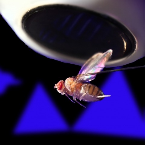
Showing 21-28 of 28 resultsDiverse sensory systems, from audition to thermosensation, feature a separation of inputs into ON (increments) and OFF (decrements) signals. In the Drosophila visual system, separate ON and OFF pathways compute the direction of motion, yet anatomical and functional studies have identified some crosstalk between these channels. We used this well-studied circuit to ask whether the motion computation depends on ON-OFF pathway crosstalk. Using whole-cell electrophysiology, we recorded visual responses of T4 (ON) and T5 (OFF) cells, mapped their composite ON-OFF receptive fields, and found that they share a similar spatiotemporal structure. We fit a biophysical model to these receptive fields that accurately predicts directionally selective T4 and T5 responses to both ON and OFF moving stimuli. This model also provides a detailed mechanistic explanation for the directional preference inversion in response to the prominent reverse-phi illusion. Finally, we used the steering responses of tethered flying flies to validate the model's predicted effects of varying stimulus parameters on the behavioral turning inversion.
A large variability in performance is observed when participants recall briefly presented lists of words. The sources of such variability are not known. Our analysis of a large data set of free recall revealed a small fraction of participants that reached an extremely high performance, including many trials with the recall of complete lists. Moreover, some of them developed a number of consistent input-position-dependent recall strategies, in particular recalling words consecutively ("chaining") or in groups of consecutively presented words ("chunking"). The time course of acquisition and particular choice of positional grouping were variable among participants. Our results show that acquiring positional strategies plays a crucial role in improvement of recall performance.
Ring attractors are a class of recurrent networks hypothesized to underlie the representation of heading direction. Such network structures, schematized as a ring of neurons whose connectivity depends on their heading preferences, can sustain a bump-like activity pattern whose location can be updated by continuous shifts along either turn direction. We recently reported that a population of fly neurons represents the animal's heading via bump-like activity dynamics. We combined two-photon calcium imaging in head-fixed flying flies with optogenetics to overwrite the existing population representation with an artificial one, which was then maintained by the circuit with naturalistic dynamics. A network with local excitation and global inhibition enforces this unique and persistent heading representation. Ring attractor networks have long been invoked in theoretical work; our study provides physiological evidence of their existence and functional architecture.
A neuron that extracts directionally selective motion information from upstream signals lacking this selectivity must compare visual responses from spatially offset inputs. Distinguishing among prevailing algorithmic models for this computation requires measuring fast neuronal activity and inhibition. In the Drosophila melanogaster visual system, a fourth-order neuron-T4-is the first cell type in the ON pathway to exhibit directionally selective signals. Here we use in vivo whole-cell recordings of T4 to show that directional selectivity originates from simple integration of spatially offset fast excitatory and slow inhibitory inputs, resulting in a suppression of responses to the nonpreferred motion direction. We constructed a passive, conductance-based model of a T4 cell that accurately predicts the neuron's response to moving stimuli. These results connect the known circuit anatomy of the motion pathway to the algorithmic mechanism by which the direction of motion is computed.
In flies, the direction of moving ON and OFF features is computed separately. T4 (ON) and T5 (OFF) are the first neurons in their respective pathways to extract a directionally selective response from their non-selective inputs. Our recent study of T4 found that the integration of offset depolarizing and hyperpolarizing inputs is critical for the generation of directional selectivity. However, T5s lack small-field inhibitory inputs, suggesting they may use a different mechanism. Here we used whole-cell recordings of T5 neurons and found a similar receptive field structure: fast depolarization and persistent, spatially offset hyperpolarization. By assaying pairwise interactions of local stimulation across the receptive field, we found no amplifying responses, only suppressive responses to the non-preferred motion direction. We then evaluated passive, biophysical models and found that a model using direct inhibition, but not the removal of excitation, can accurately predict T5 responses to a range of moving stimuli.
Hippocampal activity represents many behaviorally important variables, including context, an animal's location within a given environmental context, time, and reward. Using longitudinal calcium imaging in mice, multiple large virtual environments, and differing reward contingencies, we derived a unified probabilistic model of CA1 representations centered on a single feature-the field propensity. Each cell's propensity governs how many place fields it has per unit space, predicts its reward-related activity, and is preserved across distinct environments and over months. Propensity is broadly distributed-with many low, and some very high, propensity cells-and thus strongly shapes hippocampal representations. This results in a range of spatial codes, from sparse to dense. Propensity varied ∼10-fold between adjacent cells in salt-and-pepper fashion, indicating substantial functional differences within a presumed cell type. Intracellular recordings linked propensity to cell excitability. The stability of each cell's propensity across conditions suggests this fundamental property has anatomical, transcriptional, and/or developmental origins.
Sensory cue inputs and memory-related internal brain activities govern the firing of hippocampal neurons, but which specific firing patterns are induced by either of the two processes remains unclear. We found that sensory cues guided the firing of neurons in rats on a timescale of seconds and supported the formation of spatial firing fields. Independently of the sensory inputs, the memory-related network activity coordinated the firing of neurons not only on a second-long timescale, but also on a millisecond-long timescale, and was dependent on medial septum inputs. We propose a network mechanism that might coordinate this internally generated firing. Overall, we suggest that two independent mechanisms support the formation of spatial firing fields in hippocampus, but only the internally organized system supports short-timescale sequential firing and episodic memory.
Hippocampal place cells represent different environments with distinct neural activity patterns. Following an abrupt switch between two familiar configurations of visual cues defining two environments, the hippocampal neural activity pattern switches almost immediately to the corresponding representation. Surprisingly, during a transient period following the switch to the new environment, occasional fast transitions of activity patterns between the representations (flickering) were observed (Jezek et al. 2011). Here we show that an attractor neural network model of place cells with connections endowed with short-term synaptic plasticity can account for this phenomenon. A memory trace of the recent history of network activity is maintained in the state of the synapses, allowing the network to temporarily reactivate the representation of the previous environment in the absence of the corresponding sensory cues. The model predicts that the number of flickering events depends on the amplitude of the ongoing theta rhythm and the distance between the current position of the animal and its position at the time of cue switching. We test these predictions with new analysis of experimental data. These results suggest a potential role of short-term synaptic plasticity in recruiting the activity of different cell assemblies and in shaping hippocampal activity of behaving animals. This article is protected by copyright. All rights reserved.