Filter
Associated Lab
- Ahrens Lab (1) Apply Ahrens Lab filter
- Betzig Lab (9) Apply Betzig Lab filter
- Hess Lab (1) Apply Hess Lab filter
- Ji Lab (2) Apply Ji Lab filter
- Lavis Lab (1) Apply Lavis Lab filter
- Pedram Lab (1) Apply Pedram Lab filter
- Remove Shroff Lab filter Shroff Lab
- Wang (Shaohe) Lab (1) Apply Wang (Shaohe) Lab filter
Associated Project Team
22 Janelia Publications
Showing 11-20 of 22 resultsThe proliferation of microscopy methods for live-cell imaging offers many new possibilities for users but can also be challenging to navigate. The prevailing challenge in live-cell fluorescence microscopy is capturing intra-cellular dynamics while preserving cell viability. Computational methods can help to address this challenge and are now shifting the boundaries of what is possible to capture in living systems. In this Review, we discuss these computational methods focusing on artificial intelligence-based approaches that can be layered on top of commonly used existing microscopies as well as hybrid methods that integrate computation and microscope hardware. We specifically discuss how computational approaches can improve the signal-to-noise ratio, spatial resolution, temporal resolution and multi-colour capacity of live-cell imaging.
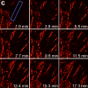
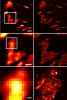
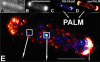
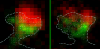
We demonstrate live-cell super-resolution imaging using photoactivated localization microscopy (PALM). The use of photon-tolerant cell lines in combination with the high resolution and molecular sensitivity of PALM permitted us to investigate the nanoscale dynamics within individual adhesion complexes (ACs) in living cells under physiological conditions for as long as 25 min, with half of the time spent collecting the PALM images at spatial resolutions down to approximately 60 nm and frame rates as short as 25 s. We visualized the formation of ACs and measured the fractional gain and loss of individual paxillin molecules as each AC evolved. By allowing observation of a wide variety of nanoscale dynamics, live-cell PALM provides insights into molecular assembly during the initiation, maturation and dissolution of cellular processes.
Commentary: The first example of true live cell and time lapse imaging by localization microscopy (as opposed to particle tracking), this paper uses the Nyquist criterion to establish a necessary condition for true spatial resolution based on the density of localized molecules – a condition often unmet in claims elsewhere in the superresolution literature.
By any method, higher spatiotemporal resolution requires increasing light exposure at the specimen, making noninvasive imaging increasingly difficult. Here, simultaneous differential interference contrast imaging is used to establish that cells behave physiologically before, during, and after PALM imaging. Similar controls are lacking from many supposed “live cell” superresolution demonstrations.
Stereocilia are unidirectional F-actin-based cylindrical protrusions on the apical surface of inner ear hair cells and function as biological mechanosensors of sound and acceleration. Development of functional stereocilia requires motor activities of unconventional myosins to transport proteins necessary for elongating the F-actin cores and to assemble the mechanoelectrical transduction (MET) channel complex. However, how each myosin localizes in stereocilia using the energy from ATP hydrolysis is only partially understood. In this study, we develop a methodology for live-cell single-molecule fluorescence microscopy of organelles protruding from the apical surface using a dual-view light-sheet microscope, diSPIM. We demonstrate that MYO7A, a component of the MET machinery, traffics as a dimer in stereocilia. Movements of MYO7A are restricted when scaffolded by the plasma membrane and F-actin as mediated by MYO7A’s interacting partners. Here, we discuss the technical details of our methodology and its future applications including analyses of cargo transportation in various organelles.
Spatial information is critical to the interrogation of developmental and tissue-level regulation of gene expression. However, this information is usually lost when global mRNA levels from tissues are measured using reverse transcriptase PCR, microarray analysis or high-throughput sequencing. By contrast, single-molecule fluorescence in situ hybridization (smFISH) preserves the spatial information of the cellular mRNA content with subcellular resolution within tissues. Here we describe an smFISH protocol that allows for the quantification of single mRNAs in Drosophila embryos, using commercially available smFISH probes (e.g., short fluorescently labeled DNA oligonucleotides) in combination with wide-field epifluorescence, confocal or instant structured illumination microscopy (iSIM, a super-resolution imaging approach) and a spot-detection algorithm. Fixed Drosophila embryos are hybridized in solution with a mixture of smFISH probes, mounted onto coverslips and imaged in 3D. Individual fluorescently labeled mRNAs are then localized within tissues and counted using spot-detection software to generate quantitative, spatially resolved gene expression data sets. With minimum guidance, a graduate student can successfully implement this protocol. The smFISH procedure described here can be completed in 4-5 d.
Recent advances in optical microscopy have enabled biological imaging beyond the diffraction limit at nanometer resolution. A general feature of most of the techniques based on photoactivated localization microscopy (PALM) or stochastic optical reconstruction microscopy (STORM) has been the use of thin biological samples in combination with total internal reflection, thus limiting the imaging depth to a fraction of an optical wavelength. However, to study whole cells or organelles that are typically up to 15 microm deep into the cell, the extension of these methods to a three-dimensional (3D) super resolution technique is required. Here, we report an advance in optical microscopy that enables imaging of protein distributions in cells with a lateral localization precision better than 50 nm at multiple imaging planes deep in biological samples. The approach is based on combining the lateral super resolution provided by PALM with two-photon temporal focusing that provides optical sectioning. We have generated super-resolution images over an axial range of approximately 10 microm in both mitochondrially labeled fixed cells, and in the membranes of living S2 Drosophila cells.
Fluorescence microscopy is an invaluable tool in biology, yet its performance is compromised when the wavefront of light is distorted due to optical imperfections or the refractile nature of the sample. Such optical aberrations can dramatically lower the information content of images by degrading image contrast, resolution, and signal. Adaptive optics (AO) methods can sense and subsequently cancel the aberrated wavefront, but are too complex, inefficient, slow, or expensive for routine adoption by most labs. Here we introduce a rapid, sensitive, and robust wavefront sensing scheme based on phase diversity, a method successfully deployed in astronomy but underused in microscopy. Our method enables accurate wavefront sensing to less than λ/35 root mean square (RMS) error with few measurements, and AO with no additional hardware besides a corrective element. After validating the method with simulations, we demonstrate calibration of a deformable mirror > 100-fold faster than comparable methods (corresponding to wavefront sensing on the ~100 ms scale), and sensing and subsequent correction of severe aberrations (RMS wavefront distortion exceeding λ/2), restoring diffraction-limited imaging on extended biological samples.
Key to understanding a protein’s biological function is the accurate determination of its spatial distribution inside a cell. Although fluorescent protein markers allow the targeting of specific proteins with molecular precision, much of this information is lost when the resultant fusion proteins are imaged with conventional, diffraction-limited optics. In response, several imaging modalities that are capable of resolution below the diffraction limit (approximately 200 nm) have emerged. Here, both single- and dual-color superresolution imaging of biological structures using photoactivated localization microscopy (PALM) are described. The examples discussed focus on adhesion complexes: dense, protein-filled assemblies that form at the interface between cells and their substrata. A particular emphasis is placed on the instrumentation and photoactivatable fluorescent protein (PA-FP) tags necessary to achieve PALM images at approximately 20 nm resolution in 5 to 30 min in fixed cells.
Commentary: A paper spearheaded by Hari which gives a thorough description of the methods and hardware needed to successfully practice PALM, including cover slip preparation, cell transfection and fixation, drift correction with fiducials, characterization of on/off contrast ratios for different photoactivted fluorescent proteins, identifying PALM-suitable cells, and mechanical and optical components of a PALM system.
Determining cell identities in imaging sequences is an important yet challenging task. The conventional method for cell identification is via cell tracking, which is complex and can be time-consuming. In this study, we propose an innovative approach to cell identification during early C. elegans embryogenesis using machine learning. We employed random forest, MLP, and LSTM models, and tested cell classification accuracy on 3D time-lapse confocal datasets spanning the first 4 hours of embryogenesis. By leveraging a small number of spatial-temporal features of individual cells, including cell trajectory and cell fate information, our models achieve an accuracy of over 90%, even with limited data. We also determine the most important feature contributions and can interpret these features in the context of biological knowledge. Our research demonstrates the success of predicting cell identities in 4D imaging sequences directly from simple spatio-temporal features.
The Escherichia coli chemotaxis network is a model system for biological signal processing. In E. coli, transmembrane receptors responsible for signal transduction assemble into large clusters containing several thousand proteins. These sensory clusters have been observed at cell poles and future division sites. Despite extensive study, it remains unclear how chemotaxis clusters form, what controls cluster size and density, and how the cellular location of clusters is robustly maintained in growing and dividing cells. Here, we use photoactivated localization microscopy (PALM) to map the cellular locations of three proteins central to bacterial chemotaxis (the Tar receptor, CheY, and CheW) with a precision of 15 nm. We find that cluster sizes are approximately exponentially distributed, with no characteristic cluster size. One-third of Tar receptors are part of smaller lateral clusters and not of the large polar clusters. Analysis of the relative cellular locations of 1.1 million individual proteins (from 326 cells) suggests that clusters form via stochastic self-assembly. The super-resolution PALM maps of E. coli receptors support the notion that stochastic self-assembly can create and maintain approximately periodic structures in biological membranes, without direct cytoskeletal involvement or active transport.
Commentary: Our goal as tool developers is to invent methods capable of uncovering new biological insights unobtainable by pre-existing technologies. A terrific example is given by this paper, where grad students Derek Greenfield and Ann McEvoy in Jan Liphardt’s group at Berkeley used our PALM to image the size and position distributions of chemotaxis proteins in E. Coli with unprecedented precision and sensitivity. Their analysis revealed that the cluster sizes follow a stretched exponential distribution, and the density of clusters is highest furthest away from the largest (e.g., polar) clusters. Both observations support a model for passive self-assembly rather than active cytoskeletal assembly of the chemotaxis network.
Within dendritic spines, actin is presumed to anchor receptors in the postsynaptic density and play numerous roles regulating synaptic transmission. However, the submicron dimensions of spines have hindered examination of actin dynamics within them and prevented live-cell discrimination of perisynaptic actin filaments. Using photoactivated localization microscopy, we measured movement of individual actin molecules within living spines. Velocity of single actin molecules along filaments, an index of filament polymerization rate, was highly heterogeneous within individual spines. Most strikingly, molecular velocity was elevated in discrete, well-separated foci occurring not principally at the spine tip, but in subdomains throughout the spine, including the neck. Whereas actin velocity on filaments at the synapse was substantially elevated, at the endocytic zone there was no enhanced polymerization activity. We conclude that actin subserves spatially diverse, independently regulated processes throughout spines. Perisynaptic actin forms a uniquely dynamic structure well suited for direct, active regulation of the synapse.
Commentary: A nice application of single particle tracking PALM (sptPALM), showing the flow of actin in the spines of live cultured neurons. Since 2008, the PALM in our lab has largely become a user facility, available to outside users as well as Janelians. Grad student Nick Frost in Tom Blanpied’s group at the U. of Maryland Med School visited on a number of occasions to use the PALM, with training and assistance from Hari.