Filter
Associated Lab
- Betzig Lab (11) Apply Betzig Lab filter
- Dudman Lab (1) Apply Dudman Lab filter
- Freeman Lab (1) Apply Freeman Lab filter
- Harris Lab (1) Apply Harris Lab filter
- Jayaraman Lab (1) Apply Jayaraman Lab filter
- Remove Ji Lab filter Ji Lab
- Keller Lab (1) Apply Keller Lab filter
- Lavis Lab (1) Apply Lavis Lab filter
- Magee Lab (2) Apply Magee Lab filter
- Shroff Lab (2) Apply Shroff Lab filter
- Svoboda Lab (2) Apply Svoboda Lab filter
Associated Project Team
Publication Date
- 2024 (1) Apply 2024 filter
- 2019 (1) Apply 2019 filter
- 2018 (5) Apply 2018 filter
- 2017 (5) Apply 2017 filter
- 2016 (4) Apply 2016 filter
- 2015 (4) Apply 2015 filter
- 2014 (3) Apply 2014 filter
- 2013 (1) Apply 2013 filter
- 2012 (2) Apply 2012 filter
- 2011 (2) Apply 2011 filter
- 2010 (1) Apply 2010 filter
- 2008 (4) Apply 2008 filter
33 Janelia Publications
Showing 11-20 of 33 resultsInherent aberrations of gradient index (GRIN) lenses used in fluorescence endomicroscopes deteriorate imaging performance. Using adaptive optics, we characterized and corrected the on-axis and off-axis aberrations of a GRIN lens with NA 0.8 at multiple focal planes. We demonstrated a rotational-transformation-based correction procedure, which enlarged the imaging area with diffraction-limited resolution with only two aberration measurements. 204.8 × 204.8 µm2 images of fluorescent beads and brain slices before and after AO corrections were obtained, with evident improvements in both image sharpness and brightness after AO correction. These results show great promises of applying adaptive optical two-photon fluorescence endomicroscope to three-dimensional (3D) imaging.
In traditional zonal wavefront sensing for adaptive optics, after local wavefront gradients are obtained, the entire wavefront can be calculated by assuming that the wavefront is a continuous surface. Such an approach will lead to sub-optimal performance in reconstructing wavefronts which are either discontinuous or undersampled by the zonal wavefront sensor. Here, we report a new method to reconstruct the wavefront by directly measuring local wavefront phases in parallel using multidither coherent optical adaptive technique. This method determines the relative phases of each pupil segment independently, and thus produces an accurate wavefront for even discontinuous wavefronts. We implemented this method in an adaptive optical two-photon fluorescence microscopy and demonstrated its superior performance in correcting large or discontinuous aberrations.
Adaptive optics by direct imaging of the wavefront distortions of a laser-induced guide star has long been used in astronomy, and more recently in microscopy to compensate for aberrations in transparent specimens. Here we extend this approach to tissues that strongly scatter visible light by exploiting the reduced scattering of near-infrared guide stars. The method enables in vivo two-photon morphological and functional imaging down to 700 μm inside the mouse brain.
Cells in the brain act as components of extended networks. Therefore, to understand neurobiological processes in a physiological context, it is essential to study them in vivo. Super-resolution microscopy has spatial resolution beyond the diffraction limit, thus promising to provide structural and functional insights that are not accessible with conventional microscopy. However, to apply it to in vivo brain imaging, we must address the challenges of 3D imaging in an optically heterogeneous tissue that is constantly in motion. We optimized image acquisition and reconstruction to combat sample motion and applied adaptive optics to correcting sample-induced optical aberrations in super-resolution structured illumination microscopy (SIM) in vivo. We imaged the brains of live zebrafish larvae and mice and observed the dynamics of dendrites and dendritic spines at nanoscale resolution.
Pulsed lasers are key elements in nonlinear bioimaging techniques such as two-photon fluorescence excitation (TPE) microscopy. Typically, however, only a percent or less of the laser power available can be delivered to the sample before photoinduced damage becomes excessive. Here we describe a passive pulse splitter that converts each laser pulse into a fixed number of sub-pulses of equal energy. We applied the splitter to TPE imaging of fixed mouse brain slices labeled with GFP and show that, in different power regimes, the splitter can be used either to increase the signal rate more than 100-fold or to reduce the rate of photobleaching by over fourfold. In living specimens, the gains were even greater: a ninefold reduction in photobleaching during in vivo imaging of Caenorhabditis elegans larvae, and a six- to 20-fold decrease in the rate of photodamage during calcium imaging of rat hippocampal brain slices.
Pulsed lasers are key elements in nonlinear bioimaging techniques such as two-photon fluorescence excitation (TPE) microscopy. Typically, however, only a percent or less of the laser power available can be delivered to the sample before photoinduced damage becomes excessive. Here we describe a passive pulse splitter that converts each laser pulse into a fixed number of sub-pulses of equal energy. We applied the splitter to TPE imaging of fixed mouse brain slices labeled with GFP and show that, in different power regimes, the splitter can be used either to increase the signal rate more than 100-fold or to reduce the rate of photobleaching by over fourfold. In living specimens, the gains were even greater: a ninefold reduction in photobleaching during in vivo imaging of Caenorhabditis elegans larvae, and a six- to 20-fold decrease in the rate of photodamage during calcium imaging of rat hippocampal brain slices.
Commentary: Na Ji came to me early in her postdoc with an idea to reduce photodamage in nonlinear microscopy by splitting the pulses from an ultrafast laser into multiple subpulses of reduced energy. In six weeks, we constructed a prototype pulse splitter and obtained initial results confirming the validity of her vision. Further experiments with Jeff Magee demonstrated that the splitter could be used to increase imaging speed or reduce photodamage in two photon microscopy by one to two orders of magnitude. This project is a great example of how quickly one can react and exploit new ideas in the Janelia environment.
The diffraction limited resolution of two photon and confocal microscope can be recovered using adaptive optics to explore the detailed neuronal network in the brains of zebrafish and mouse in vivo.
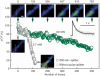
In vivo calcium imaging from axons provides direct interrogation of afferent neural activity, informing the neural representations that a local circuit receives. Unlike in somata and dendrites, axonal recording of neural activity-both electrically and optically-has been difficult to achieve, thus preventing comprehensive understanding of neuronal circuit function. Here we developed an active transportation strategy to enrich GCaMP6, a genetically encoded calcium indicator, uniformly in axons with sufficient brightness, signal-to-noise ratio, and photostability to allow robust, structure-specific imaging of presynaptic activity in awake mice. Axon-targeted GCaMP6 enables frame-to-frame correlation for motion correction in axons and permits subcellular-resolution recording of axonal activity in previously inaccessible deep-brain areas. We used axon-targeted GCaMP6 to record layer-specific local afferents without contamination from somata or from intermingled dendrites in the cortex. We expect that axon-targeted GCaMP6 will facilitate new applications in investigating afferent signals relayed by genetically defined neuronal populations within and across specific brain regions.
Hyperspectral stimulated Raman scattering microscopy is deployed to measure single-membrane vibrational spectrum as a function of membrane potential. Using erythrocyte ghost as a model, quantitative correlation between transmembrane potential and Raman spectral profile was found. Specifically, the ratio between the area under Raman band at ∼2930 cm−1 and that at ∼2850 cm−1 increased by ∼2.6 times when the potential across the erythrocyte ghost membrane varied from +10 mV to −10 mV. Our results show the feasibility of employing stimulated Raman scattering microscopy to probe the membrane potential without labeling.
The ability to image neurons anywhere in the mammalian brain is a major goal of optical microscopy. Here we describe a minimally invasive microendoscopy system for studying the morphology and function of neurons at depth. Utilizing a guide cannula with an ultrathin wall, we demonstrated in vivo two-photon fluorescence imaging of deeply buried nuclei such as the striatum (2.5 mm depth), substantia nigra (4.4 mm depth) and lateral hypothalamus (5.0 mm depth) in mouse brain. We reported, for the first time, the observation of neuronal activity with subcellular resolution in the lateral hypothalamus and substantia nigra of head-fixed awake mice.