Filter
Associated Lab
- Baker Lab (2) Apply Baker Lab filter
- Betzig Lab (3) Apply Betzig Lab filter
- Cardona Lab (2) Apply Cardona Lab filter
- Chklovskii Lab (1) Apply Chklovskii Lab filter
- Druckmann Lab (1) Apply Druckmann Lab filter
- Eddy/Rivas Lab (3) Apply Eddy/Rivas Lab filter
- Fetter Lab (2) Apply Fetter Lab filter
- Harris Lab (1) Apply Harris Lab filter
- Hess Lab (2) Apply Hess Lab filter
- Jayaraman Lab (2) Apply Jayaraman Lab filter
- Ji Lab (1) Apply Ji Lab filter
- Keller Lab (2) Apply Keller Lab filter
- Looger Lab (6) Apply Looger Lab filter
- Magee Lab (3) Apply Magee Lab filter
- Reiser Lab (2) Apply Reiser Lab filter
- Riddiford Lab (2) Apply Riddiford Lab filter
- Rubin Lab (2) Apply Rubin Lab filter
- Saalfeld Lab (1) Apply Saalfeld Lab filter
- Scheffer Lab (3) Apply Scheffer Lab filter
- Shroff Lab (1) Apply Shroff Lab filter
- Simpson Lab (3) Apply Simpson Lab filter
- Sternson Lab (1) Apply Sternson Lab filter
- Svoboda Lab (7) Apply Svoboda Lab filter
- Truman Lab (4) Apply Truman Lab filter
Associated Project Team
Associated Support Team
Publication Date
- December 2010 (3) Apply December 2010 filter
- November 2010 (4) Apply November 2010 filter
- October 2010 (5) Apply October 2010 filter
- September 2010 (3) Apply September 2010 filter
- August 2010 (7) Apply August 2010 filter
- July 2010 (2) Apply July 2010 filter
- June 2010 (6) Apply June 2010 filter
- May 2010 (3) Apply May 2010 filter
- April 2010 (5) Apply April 2010 filter
- March 2010 (1) Apply March 2010 filter
- February 2010 (6) Apply February 2010 filter
- January 2010 (16) Apply January 2010 filter
- Remove 2010 filter 2010
61 Janelia Publications
Showing 41-50 of 61 resultsIt is well known that light-sheet illumination can enable optically sectioned wide-field imaging of macroscopic samples. However, the optical sectioning capacity of a light-sheet macroscope is undermined by sample-induced scattering or aberrations that broaden the thickness of the sheet illumination. We present a technique to enhance the optical sectioning capacity of a scanning light-sheet microscope by out-of-focus background rejection. The technique, called HiLo microscopy, makes use of two images sequentially acquired with uniform and structured sheet illumination. An optically sectioned image is then synthesized by fusing high and low spatial frequency information from both images. The benefits of combining light-sheet macroscopy and HiLo background rejection are demonstrated in optically cleared whole mouse brain samples, using both green fluorescent protein (GFP)-fluorescence and dark-field scattered light contrast.
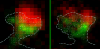
Automatic alignment (registration) of 3D images of adult fruit fly brains is often influenced by the significant displacement of the relative locations of the two optic lobes (OLs) and the center brain (CB). In one of our ongoing efforts to produce a better image alignment pipeline of adult fruit fly brains, we consider separating CB and OLs and align them independently. This paper reports our automatic method to segregate CB and OLs, in particular under conditions where the signal to noise ratio (SNR) is low, the variation of the image intensity is big, and the relative displacement of OLs and CB is substantial. We design an algorithm to find a minimum-cost 3D surface in a 3D image stack to best separate an OL (of one side, either left or right) from CB. This surface is defined as an aggregation of the respective minimum-cost curves detected in each individual 2D image slice. Each curve is defined by a list of control points that best segregate OL and CB. To obtain the locations of these control points, we derive an energy function that includes an image energy term defined by local pixel intensities and two internal energy terms that constrain the curve’s smoothness and length. Gradient descent method is used to optimize this energy function. To improve both the speed and robustness of the method, for each stack, the locations of optimized control points in a slice are taken as the initialization prior for the next slice. We have tested this approach on simulated and real 3D fly brain image stacks and demonstrated that this method can reasonably segregate OLs from CBs despite the aforementioned difficulties.
Reconstructing neuronal circuits at the level of synapses is a central problem in neuroscience, and the focus of the nascent field of connectomics. Previously used to reconstruct the C. elegans wiring diagram, serial-section transmission electron microscopy (ssTEM) is a proven technique for the task. However, to reconstruct more complex circuits, ssTEM will require the automation of image processing. We review progress in the processing of electron microscopy images and, in particular, a semi-automated reconstruction pipeline deployed at Janelia. Drosophila circuits underlying identified behaviors are being reconstructed in the pipeline with the goal of generating a complete Drosophila connectome.
The Drosophila melanogaster sex hierarchy controls sexual differentiation of somatic cells via the activities of the terminal genes in the hierarchy, doublesex (dsx) and fruitless (fru). We have targeted an insertion of GAL4 into the dsx gene, allowing us to visualize dsx-expressing cells in both sexes. Developmentally and as adults, we find that both XX and XY individuals are fine mosaics of cells and tissues that express dsx and/or fruitless (fru(M)), and hence have the potential to sexually differentiate, and those that don’t. Evolutionary considerations suggest such a mosaic expression of sexuality is likely to be a property of other animal species having two sexes. These results have also led to a major revision of our view of how sex-specific functions are regulated by the sex hierarchy in flies. Rather than there being a single regulatory event that governs the activities of all downstream sex determination regulatory genes-turning on Sex lethal (Sxl) RNA splicing activity in females while leaving it turned off in males-there are, in addition, elaborate temporal and spatial transcriptional controls on the expression of the terminal regulatory genes, dsx and fru. Thus tissue-specific aspects of sexual development are jointly specified by post-transcriptional control by Sxl and by the transcriptional controls of dsx and fru expression.
Within dendritic spines, actin is presumed to anchor receptors in the postsynaptic density and play numerous roles regulating synaptic transmission. However, the submicron dimensions of spines have hindered examination of actin dynamics within them and prevented live-cell discrimination of perisynaptic actin filaments. Using photoactivated localization microscopy, we measured movement of individual actin molecules within living spines. Velocity of single actin molecules along filaments, an index of filament polymerization rate, was highly heterogeneous within individual spines. Most strikingly, molecular velocity was elevated in discrete, well-separated foci occurring not principally at the spine tip, but in subdomains throughout the spine, including the neck. Whereas actin velocity on filaments at the synapse was substantially elevated, at the endocytic zone there was no enhanced polymerization activity. We conclude that actin subserves spatially diverse, independently regulated processes throughout spines. Perisynaptic actin forms a uniquely dynamic structure well suited for direct, active regulation of the synapse.
Commentary: A nice application of single particle tracking PALM (sptPALM), showing the flow of actin in the spines of live cultured neurons. Since 2008, the PALM in our lab has largely become a user facility, available to outside users as well as Janelians. Grad student Nick Frost in Tom Blanpied’s group at the U. of Maryland Med School visited on a number of occasions to use the PALM, with training and assistance from Hari.
The stabilization of new spines in the barrel cortex is enhanced after whisker trimming, but its relationship to experience-dependent plasticity is unclear. Here we show that in wild-type mice, whisker potentiation and spine stabilization are most pronounced for layer 5 neurons at the border between spared and deprived barrel columns. In homozygote alphaCaMKII-T286A mice, which lack experience-dependent potentiation of responses to spared whiskers, there is no increase in new spine stabilization at the border between barrel columns after whisker trimming. Our data provide a causal link between new spine synapses and plasticity of adult cortical circuits and suggest that alphaCaMKII autophosphorylation plays a role in the stabilization but not formation of new spines.
BACKGROUND: Previous studies suggest that mechanical feedback could coordinate morphogenetic events in embryos. Furthermore, embryonic tissues have complex structure and composition and undergo large deformations during morphogenesis. Hence we expect highly non-linear and loading-rate dependent tissue mechanical properties in embryos. METHODOLOGY/PRINCIPAL FINDINGS: We used micro-aspiration to test whether a simple linear viscoelastic model was sufficient to describe the mechanical behavior of gastrula stage Xenopus laevis embryonic tissue in vivo. We tested whether these embryonic tissues change their mechanical properties in response to mechanical stimuli but found no evidence of changes in the viscoelastic properties of the tissue in response to stress or stress application rate. We used this model to test hypotheses about the pattern of force generation during electrically induced tissue contractions. The dependence of contractions on suction pressure was most consistent with apical tension, and was inconsistent with isotropic contraction. Finally, stiffer clutches generated stronger contractions, suggesting that force generation and stiffness may be coupled in the embryo. CONCLUSIONS/SIGNIFICANCE: The mechanical behavior of a complex, active embryonic tissue can be surprisingly well described by a simple linear viscoelastic model with power law creep compliance, even at high deformations. We found no evidence of mechanical feedback in this system. Together these results show that very simple mechanical models can be useful in describing embryo mechanics.
BACKGROUND: Conditional gene activation is an efficient strategy for studying gene function in genetically modified animals. Among the presently available gene switches, the tetracycline-regulated system has attracted considerable interest because of its unique potential for reversible and adjustable gene regulation. RESULTS: To investigate whether the ubiquitously expressed Gt(ROSA)26Sor locus enables uniform DOX-controlled gene expression, we inserted the improved tetracycline-regulated transcription activator iM2 together with an iM2 dependent GFP gene into the Gt(ROSA)26Sor locus, using gene targeting to generate ROSA26-iM2-GFP (R26t1Δ) mice. Despite the presence of ROSA26 promoter driven iM2, R26t1Δ mice showed very sparse DOX-activated expression of different iM2-responsive reporter genes in the brain, mosaic expression in peripheral tissues and more prominent expression in erythroid, myeloid and lymphoid lineages, in hematopoietic stem and progenitor cells and in olfactory neurons. CONCLUSIONS: The finding that gene regulation by the DOX-activated transcriptional factor iM2 in the Gt(ROSA)26Sor locus has its limitations is of importance for future experimental strategies involving transgene activation from the endogenous ROSA26 promoter. Furthermore, our ROSA26-iM2 knock-in mouse model (R26t1Δ) represents a useful tool for implementing gene function in vivo especially under circumstances requiring the side-by-side comparison of gene manipulated and wild type cells. Since the ROSA26-iM2 mouse allows mosaic gene activation in peripheral tissues and haematopoietic cells, this model will be very useful for uncovering previously unknown or unsuspected phenotypes.
The primary auditory cortex (A1) is organized tonotopically, with neurons sensitive to high and low frequencies arranged in a rostro-caudal gradient. We used laser scanning photostimulation in acute slices to study the organization of local excitatory connections onto layers 2 and 3 (L2/3) of the mouse A1. Consistent with the organization of other cortical regions, synaptic inputs along the isofrequency axis (orthogonal to the tonotopic axis) arose predominantly within a column. By contrast, we found that local connections along the tonotopic axis differed from those along the isofrequency axis: some input pathways to L3 (but not L2) arose predominantly out-of-column. In vivo cell-attached recordings revealed differences between the sound-responsiveness of neurons in L2 and L3. Our results are consistent with the hypothesis that auditory cortical microcircuitry is specialized to the one-dimensional representation of frequency in the auditory cortex.