Filter
Associated Lab
- Ahrens Lab (3) Apply Ahrens Lab filter
- Harris Lab (4) Apply Harris Lab filter
- Hermundstad Lab (1) Apply Hermundstad Lab filter
- Jayaraman Lab (7) Apply Jayaraman Lab filter
- Karpova Lab (1) Apply Karpova Lab filter
- Leonardo Lab (2) Apply Leonardo Lab filter
- Remove Looger Lab filter Looger Lab
- Podgorski Lab (2) Apply Podgorski Lab filter
- Rubin Lab (1) Apply Rubin Lab filter
- Schreiter Lab (13) Apply Schreiter Lab filter
- Svoboda Lab (10) Apply Svoboda Lab filter
- Tillberg Lab (1) Apply Tillberg Lab filter
- Turner Lab (1) Apply Turner Lab filter
- Zlatic Lab (1) Apply Zlatic Lab filter
Associated Project Team
Associated Support Team
16 Janelia Publications
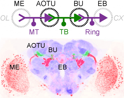
Showing 11-16 of 16 resultsMany animals orient using visual cues, but how a single cue is selected from among many is poorly understood. Here we show that Drosophila ring neurons—central brain neurons implicated in navigation—display visual stimulus selection. Using in vivo two-color two-photon imaging with genetically encoded calcium indicators, we demonstrate that individual ring neurons inherit simple-cell-like receptive fields from their upstream partners. Stimuli in the contralateral visual field suppressed responses to ipsilateral stimuli in both populations. Suppression strength depended on when and where the contralateral stimulus was presented, an effect stronger in ring neurons than in their upstream inputs. This history-dependent effect on the temporal structure of visual responses, which was well modeled by a simple biphasic filter, may determine how visual references are selected for the fly's internal compass. Our approach highlights how two-color calcium imaging can help identify and localize the origins of sensory transformations across synaptically connected neural populations.
Genetically encoded calcium indicators (GECIs) are powerful tools for systems neuroscience. Recent efforts in protein engineering have significantly increased the performance of GECIs. The state-of-the art single-wavelength GECI, GCaMP3, has been deployed in a number of model organisms and can reliably detect three or more action potentials in short bursts in several systems in vivo . Through protein structure determination, targeted mutagenesis, high-throughput screening, and a battery of in vitro assays, we have increased the dynamic range of GCaMP3 by severalfold, creating a family of “GCaMP5” sensors. We tested GCaMP5s in several systems: cultured neurons and astrocytes, mouse retina, and in vivo in Caenorhabditis chemosensory neurons, Drosophila larval neuromuscular junction and adult antennal lobe, zebrafish retina and tectum, and mouse visual cortex. Signal-to-noise ratio was improved by at least 2- to 3-fold. In the visual cortex, two GCaMP5 variants detected twice as many visual stimulus-responsive cells as GCaMP3. By combining in vivo imaging with electrophysiology we show that GCaMP5 fluorescence provides a more reliable measure of neuronal activity than its predecessor GCaMP3.GCaMP5allows more sensitive detection of neural activity in vivo andmayfind widespread applications for cellular imaging in general.
Genetically encoded calcium indicators (GECIs) allow measurement of activity in large populations of neurons and in small neuronal compartments, over times of milliseconds to months. Although GFP-based GECIs are widely used for in vivo neurophysiology, GECIs with red-shifted excitation and emission spectra have advantages for in vivo imaging because of reduced scattering and absorption in tissue, and a consequent reduction in phototoxicity. However, current red GECIs are inferior to the state-of-the-art GFP-based GCaMP6 indicators for detecting and quantifying neural activity. Here we present improved red GECIs based on mRuby (jRCaMP1a, b) and mApple (jRGECO1a), with sensitivity comparable to GCaMP6. We characterized the performance of the new red GECIs in cultured neurons and in mouse, Drosophila, zebrafish and C. elegans in vivo. Red GECIs facilitate deep-tissue imaging, dual-color imaging together with GFP-based reporters, and the use of optogenetics in combination with calcium imaging.
Genetically-encoded calcium indicators (GECIs) facilitate imaging activity of genetically defined neuronal populations in vivo. The high intracellular GECI concentrations required for in vivo imaging are usually achieved by viral gene transfer using adeno-associated viruses. Transgenic expression of GECIs promises important advantages, including homogeneous, repeatable, and stable expression without the need for invasive virus injections. Here we present the generation and characterization of transgenic mice expressing the GECIs GCaMP6s or GCaMP6f under the Thy1 promoter. We quantified GCaMP6 expression across brain regions and neurons and compared to other transgenic mice and AAV-mediated expression. We tested three mouse lines for imaging in the visual cortex in vivo and compared their performance to mice injected with AAV expressing GCaMP6. Furthermore, we show that GCaMP6 Thy1 transgenic mice are useful for long-term, high-sensitivity imaging in behaving mice.
Fluorescent calcium sensors are widely used to image neural activity. Using structure-based mutagenesis and neuron-based screening, we developed a family of ultrasensitive protein calcium sensors (GCaMP6) that outperformed other sensors in cultured neurons and in zebrafish, flies and mice in vivo. In layer 2/3 pyramidal neurons of the mouse visual cortex, GCaMP6 reliably detected single action potentials in neuronal somata and orientation-tuned synaptic calcium transients in individual dendritic spines. The orientation tuning of structurally persistent spines was largely stable over timescales of weeks. Orientation tuning averaged across spine populations predicted the tuning of their parent cell. Although the somata of GABAergic neurons showed little orientation tuning, their dendrites included highly tuned dendritic segments (5–40-µm long). GCaMP6 sensors thus provide new windows into the organization and dynamics of neural circuits over multiple spatial and temporal scales.
Mechanisms that entrain and drive rhythmic epileptiform discharges remain debated. Traditionally, this quest has been focusing on interneuronal networks driven by GABAergic connections that activate synaptic or extrasynaptic receptors. However, synchronised interneuronal discharges could also trigger a transient elevation of extracellular GABA across the tissue volume, thus raising tonic GABAA receptor conductance (Gtonic) in multiple cells. Here, we use patch-clamp GABA ‘sniffer’ and optical GABA sensor to show that periodic epileptiform discharges are preceded by region-wide, rising waves of extracellular GABA. Neural network simulations that incorporate volume-transmitted GABA signals point to mechanistic principles underpinning this relationship. We validate this hypothesis using simultaneous patch-clamp recordings from multiple nerve cells, selective optogenetic stimulation of fast-spiking interneurons. Critically, we manipulate GABA uptake to suppress extracellular GABA waves but not synaptic GABAergic transmission, which shows a clear effect on rhythm generation. Our findings thus unveil a key role of extrasynaptic, volume-transmitted GABA actions in pacing regenerative rhythmic activity in brain networks.

