Filter
Associated Lab
- Ahrens Lab (7) Apply Ahrens Lab filter
- Druckmann Lab (2) Apply Druckmann Lab filter
- Dudman Lab (1) Apply Dudman Lab filter
- Freeman Lab (2) Apply Freeman Lab filter
- Harris Lab (7) Apply Harris Lab filter
- Hermundstad Lab (1) Apply Hermundstad Lab filter
- Hess Lab (2) Apply Hess Lab filter
- Jayaraman Lab (10) Apply Jayaraman Lab filter
- Karpova Lab (2) Apply Karpova Lab filter
- Keller Lab (5) Apply Keller Lab filter
- Lavis Lab (8) Apply Lavis Lab filter
- Leonardo Lab (4) Apply Leonardo Lab filter
- Remove Looger Lab filter Looger Lab
- Podgorski Lab (6) Apply Podgorski Lab filter
- Rubin Lab (2) Apply Rubin Lab filter
- Schreiter Lab (24) Apply Schreiter Lab filter
- Simpson Lab (1) Apply Simpson Lab filter
- Spruston Lab (1) Apply Spruston Lab filter
- Sternson Lab (2) Apply Sternson Lab filter
- Svoboda Lab (20) Apply Svoboda Lab filter
- Tervo Lab (1) Apply Tervo Lab filter
- Tillberg Lab (1) Apply Tillberg Lab filter
- Turner Lab (1) Apply Turner Lab filter
- Zlatic Lab (1) Apply Zlatic Lab filter
Associated Project Team
Associated Support Team
- Anatomy and Histology (2) Apply Anatomy and Histology filter
- Cryo-Electron Microscopy (1) Apply Cryo-Electron Microscopy filter
- Electron Microscopy (1) Apply Electron Microscopy filter
- Fly Facility (1) Apply Fly Facility filter
- Janelia Experimental Technology (2) Apply Janelia Experimental Technology filter
- Molecular Genomics (1) Apply Molecular Genomics filter
- Primary & iPS Cell Culture (2) Apply Primary & iPS Cell Culture filter
- Quantitative Genomics (2) Apply Quantitative Genomics filter
- Scientific Computing Software (1) Apply Scientific Computing Software filter
- Viral Tools (1) Apply Viral Tools filter
- Vivarium (1) Apply Vivarium filter
Publication Date
- 2024 (1) Apply 2024 filter
- 2023 (5) Apply 2023 filter
- 2022 (7) Apply 2022 filter
- 2021 (11) Apply 2021 filter
- 2020 (7) Apply 2020 filter
- 2019 (15) Apply 2019 filter
- 2018 (8) Apply 2018 filter
- 2017 (6) Apply 2017 filter
- 2016 (10) Apply 2016 filter
- 2015 (9) Apply 2015 filter
- 2014 (11) Apply 2014 filter
- 2013 (10) Apply 2013 filter
- 2012 (13) Apply 2012 filter
- 2011 (7) Apply 2011 filter
- 2010 (6) Apply 2010 filter
- 2009 (7) Apply 2009 filter
- 2008 (3) Apply 2008 filter
136 Janelia Publications
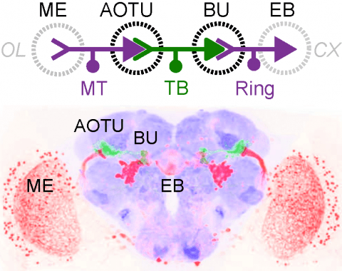
Showing 101-110 of 136 resultsMany animals orient using visual cues, but how a single cue is selected from among many is poorly understood. Here we show that Drosophila ring neurons—central brain neurons implicated in navigation—display visual stimulus selection. Using in vivo two-color two-photon imaging with genetically encoded calcium indicators, we demonstrate that individual ring neurons inherit simple-cell-like receptive fields from their upstream partners. Stimuli in the contralateral visual field suppressed responses to ipsilateral stimuli in both populations. Suppression strength depended on when and where the contralateral stimulus was presented, an effect stronger in ring neurons than in their upstream inputs. This history-dependent effect on the temporal structure of visual responses, which was well modeled by a simple biphasic filter, may determine how visual references are selected for the fly's internal compass. Our approach highlights how two-color calcium imaging can help identify and localize the origins of sensory transformations across synaptically connected neural populations.
Genetically encoded calcium indicators (GECIs) are powerful tools for systems neuroscience. Recent efforts in protein engineering have significantly increased the performance of GECIs. The state-of-the art single-wavelength GECI, GCaMP3, has been deployed in a number of model organisms and can reliably detect three or more action potentials in short bursts in several systems in vivo . Through protein structure determination, targeted mutagenesis, high-throughput screening, and a battery of in vitro assays, we have increased the dynamic range of GCaMP3 by severalfold, creating a family of “GCaMP5” sensors. We tested GCaMP5s in several systems: cultured neurons and astrocytes, mouse retina, and in vivo in Caenorhabditis chemosensory neurons, Drosophila larval neuromuscular junction and adult antennal lobe, zebrafish retina and tectum, and mouse visual cortex. Signal-to-noise ratio was improved by at least 2- to 3-fold. In the visual cortex, two GCaMP5 variants detected twice as many visual stimulus-responsive cells as GCaMP3. By combining in vivo imaging with electrophysiology we show that GCaMP5 fluorescence provides a more reliable measure of neuronal activity than its predecessor GCaMP3.GCaMP5allows more sensitive detection of neural activity in vivo andmayfind widespread applications for cellular imaging in general.
Correlative light and electron microscopy (CLEM) combines the power of electron microscopy, with its excellent resolution and contrast, with that of fluorescence imaging, which allows the staining of specific molecules, organelles, and cell populations. Fluorescence imaging is also readily compatible with live cells and behaving animals, facilitating real-time visualization of cellular processes, potentially followed by electron microscopic reconstruction. Super-resolution single-molecule localization microscopy is a relatively new modality that harnesses the ability of some fluorophores to photoconvert, through which localization precision better than Abbe’s diffraction limit is achieved through iterative high-resolution localization of single-molecule emitters. Here we describe our lab’s recent progress in the development of reagents and techniques for super-resolution single-molecule localization CLEM and their applications to biological problems.
Light-inducible dimerization protein modules enable precise temporal and spatial control of biological processes in non-invasive fashion. Among them, Magnets are small modules engineered from the photoreceptor Vivid by orthogonalizing the homodimerization interface into complementary heterodimers. Both Magnets components, which are well-tolerated as protein fusion partners, are photoreceptors requiring simultaneous photoactivation to interact, enabling high spatiotemporal confinement of dimerization with a single-excitation wavelength. However, Magnets require concatemerization for efficient responses and cell preincubation at 28C to be functional. Here we overcome these limitations by engineering an optimized Magnets pair requiring neither concatemerization nor low temperature preincubation. We validated these 'enhanced' Magnets (eMags) by using them to rapidly and reversibly recruit proteins to subcellular organelles, to induce organelle contacts, and to reconstitute OSBP-VAP ER-Golgi tethering implicated in phosphatidylinositol-4-phosphate transport and metabolism. eMags represent a very effective tool to optogenetically manipulate physiological processes over whole cells or in small subcellular volumes.
Nociception generally evokes rapid withdrawal behavior in order to protect the tissue from harmful insults. Most nociceptive neurons responding to mechanical insults display highly branched dendrites, an anatomy shared by Caenorhabditis elegans FLP and PVD neurons, which mediate harsh touch responses. Although several primary molecular nociceptive sensors have been characterized, less is known about modulation and amplification of noxious signals within nociceptor neurons. First, we analyzed the FLP/PVD network by optogenetics and studied integration of signals from these cells in downstream interneurons. Second, we investigated which genes modulate PVD function, based on prior single-neuron mRNA profiling of PVD.
To truly understand biological systems, one must possess the ability to selectively manipulate their parts and observe the outcome. (For purposes of this review, we refer mostly to targets of neuroscience; however, the principles covered here largely extend to myriad samples from microbes to plants to the intestine, etc.). Drugs are the most commonly employed way of introducing such perturbations, but they act on endogenous proteins that frequently exist in multiple cell types, complicating the interpretation of experiments. Whatever the applied stimulus, it is best to introduce optimized exogenous reagents into the systems under studydenabling manipulations to be targeted to speci!c cells and pathways. (It is also possible to target manipulations through other means, such as drugs that acquire cell-type speci!city through targeting via antibodies and/or cell surface receptor ligands, but as far as we are aware, existing reagents fall short in terms of necessary speci!city.) Many types of perturbations are useful in living systems and can be divided into rough categories such as the following: depolarize or hyperpolarize cells, induce or repress the activity of a speci!c pathway, induce or inhibit expression of a particular gene, activate or repress a speci!c protein, degrade a speci!c protein, etc. User-supplied triggers for such manipulations to occur include the following: addition of a small molecule (“chemogenetics”dideally inert on endogenous proteins) [1], sound waves (“sonogenetics”) [2], alteration of temperature (“thermogenetics”d almost exclusively used for small invertebrates) [3], and light (“optogenetics”). There are reports of using magnetic !elds (“magnetogenetics”) [4], but there is no evidence that such effects are reproducible or even physically possible [5,6]. Of these, the most commonly used, for multiple reasons, is light. Many factors make light an ideal user-controlled stimulus for the manipulation of samples. Light is quickly delivered, and most light-sensitive proteins and other molecules respond quickly to light stimuli, making many optogenetic systems relatively rapid in comparison to, for instance, drug-modulated systems. Light is also quite easy to deliver in localized patterns, allowing for targeted stimulation. Multiple wavelengths can be delivered separately to distinct (or overlapping) regions, potentially allowing combinatorial control of diverse components. Finally, light can be delivered to shallow brain regions (and peripheral sites) relatively noninvasively, and to deeper brain regions with some effort. However, there are also a number of shortcomings of using light for control. Robust and uniform penetration of light into the sample is the most signi!cant concern. For systems requiring modulation of many cells, particularly at depth, the use of systems controlled by small molecule drugs would generally be recommended instead of optogenetic approaches. When light is delivered through the use of !bers, lenses, or other optical devices, such interventions can produce signi!- cant cellular death, scar formation, and biofouling. The foreign-body response of tissue to objects triggers substantial molecular alterations, the implications of which are incompletely de!ned, but can involve reactive astrogliosis, oxidative stress, and perturbed vascularization. Head-mounted lightdelivery devices can be heavy and/or restrictive, and thus perturb behavior, particularly for small animals (e.g., mouse behavior is much more disrupted than rat behavior). More generally, all light causes tissue heating, which can have dramatic effects on cell health, physiology, and animal behavior. This is most concerning for tiny animals such as "ies. Light itself also damages tissue, most obviously through photochemistry (e.g., oxidation and radicalization) and photobleaching of critical endogenousmolecules. Furthermore, of course, light is ubiquitous, meaning that the sample is never completely unstimulated, despite precautions. Light passes through the eyes into the brain with surprising ease, and even through the skull with modest ef!cacy [7]dwhich can disrupt animal behavior (as can the converse: stimulating light in the brain perceived as a visual stimulus through the back of the eyes.) Light-responsive proteins exist in all samples, particularly in the eyes but to some extent in all tissuesdnotably, deep-brain photoreceptors [8]. The use of optogenetic tools has accelerated research on many fronts in disparate !elds. Additional, perhaps most, limitations on the utility of optogenetics must, however, be placed squarely on the shortcomings of the current suite of tools (and potential inherent limits in their performance.) The vast majority of optogenetic effectors are gated by blue light, which has signi!cant penetration issues and can be phototoxic under high intensity; redder wavelengths would in general be preferred. Furthermore, multiplexing requires tools making use of other parts of the visible spectrum (and redder wavelengths). A related issue is that most chromophores for optogenetic reagents have very broad action spectra (w250 nm bandwidth for retinal; w200 nm bandwidth for "avin), complicating both multiplexing and their use alongside many optical imaging reagentsdnarrower action spectra would be preferred for effectors in most situations. More generally, the current classes of optogenetic effectors are few, mostly limited to (1) channels and pumps (most with poor ion selectivity), (2) dimerizers, and (3) a handful of enzymes. The number of optogenetic tools that perform a very speci!c function in cells is small. Although progress has undeniably been made, much additional research and engineering will be required to dramatically expand the optogenetic toolkit. Rather than providing a survey of research !ndings, this review covers general considerations of optogenetics experiments, and then focuses largely on molecular tools: the existing suite, their features and limitations, and goals for the creation and validation of additional reagents.
Fold-switching proteins challenge the one-sequence-one-structure paradigm by adopting multiple stable folds. Nevertheless, it is uncertain whether fold switchers are naturally pervasive or rare exceptions to the well-established rule. To address this question, we developed a predictive method and applied it to the NusG superfamily of >15,000 transcription factors. We predicted that a substantial population (25%) of the proteins in this family switch folds. Circular dichroism and nuclear magnetic resonance spectroscopies of 10 sequence-diverse variants confirmed our predictions. Thus, we leveraged family-wide predictions to determine both conserved contacts and taxonomic distributions of fold-switching proteins. Our results indicate that fold switching is pervasive in the NusG superfamily and that the single-fold paradigm significantly biases structure-prediction strategies.
Glucose is an essential source of energy for the brain. Recently, the development of genetically encoded fluorescent biosensors has allowed real time visualization of glucose dynamics from individual neurons and astrocytes. A major difficulty for this approach, even for ratiometric sensors, is the lack of a practical method to convert such measurements into actual concentrations in ex vivo brain tissue or in vivo. Fluorescence lifetime imaging provides a strategy to overcome this. In a previous study, we reported the lifetime glucose sensor iGlucoSnFR-TS (then called SweetieTS) for monitoring changes in neuronal glucose levels in response to stimulation. This genetically encoded sensor was generated by combining the Thermus thermophilus glucose-binding protein with a circularly permuted variant of the monomeric fluorescent protein T-Sapphire. Here, we provide more details on iGlucoSnFR-TS design and characterization, as well as pH and temperature sensitivities. For accurate estimation of glucose concentrations, the sensor must be calibrated at the same temperature as the experiments. We find that when the extracellular glucose concentration is in the range 2-10 mM, the intracellular glucose concentration in hippocampal neurons from acute brain slices is ~20% of the nominal external glucose concentration (~0.4-2 mM). We also measured the cytosolic neuronal glucose concentration in vivo, finding a range of ~0.7-2.5 mM in cortical neurons from awake mice.
Although mRNA translation is a fundamental biological process, it has never been imaged in real-time with single molecule precision in vivo. To achieve this, we developed Nascent Chain Tracking (NCT), a technique that uses multi-epitope tags and antibody-based fluorescent probes to quantify single mRNA protein synthesis dynamics. NCT reveals an elongation rate of ~10 amino acids per second, with initiation occurring stochastically every ~30 s. Polysomes contain ~1 ribosome every 200-900 nucleotides and are globular rather than elongated in shape. By developing multi-color probes, we show most polysomes act independently; however, a small fraction (~5%) form complexes in which two distinct mRNAs can be translated simultaneously. The sensitivity and versatility of NCT make it a powerful new tool for quantifying mRNA translation kinetics.
Retinal bipolar cells (BCs) transmit visual signals in parallel channels from the outer to the inner retina, where they provide glutamatergic inputs to specific networks of amacrine and ganglion cells. Intricate network computation at BC axon terminals has been proposed as a mechanism for complex network computation, such as direction selectivity, but direct knowledge of the receptive field property and the synaptic connectivity of the axon terminals of various BC types is required in order to understand the role of axonal computation by BCs. The present study tested the essential assumptions of the presynaptic model of direction selectivity at axon terminals of three functionally distinct BC types that ramify in the direction-selective strata of the mouse retina. Results from two-photon Ca2+ imaging, optogenetic stimulation, and dual patch-clamp recording demonstrated that (1) CB5 cells do not receive fast GABAergic synaptic feedback from starburst amacrine cells (SACs), (2) light-evoked and spontaneous Ca2+ responses are well coordinated among various local regions of CB5 axon terminals, (3) CB5 axon terminals are not directionally selective, (4) CB5 cells consist of two novel functional subtypes with distinct receptive field structures, (5) CB7 cells provide direct excitatory synaptic inputs to, but receive no direct GABAergic synaptic feedback from SACs, and (6) CB7 axon terminals are not directionally selective either. These findings help to simplify models of direction selectivity by ruling out complex computation at BC terminals. They also show that CB5 comprises two functional subclasses of BCs.

