Filter
Associated Lab
- Aso Lab (2) Apply Aso Lab filter
- Baker Lab (2) Apply Baker Lab filter
- Betzig Lab (5) Apply Betzig Lab filter
- Bock Lab (1) Apply Bock Lab filter
- Branson Lab (3) Apply Branson Lab filter
- Card Lab (1) Apply Card Lab filter
- Cardona Lab (1) Apply Cardona Lab filter
- Chklovskii Lab (1) Apply Chklovskii Lab filter
- Cui Lab (6) Apply Cui Lab filter
- Druckmann Lab (2) Apply Druckmann Lab filter
- Eddy/Rivas Lab (1) Apply Eddy/Rivas Lab filter
- Fetter Lab (1) Apply Fetter Lab filter
- Gonen Lab (4) Apply Gonen Lab filter
- Harris Lab (3) Apply Harris Lab filter
- Heberlein Lab (1) Apply Heberlein Lab filter
- Hess Lab (3) Apply Hess Lab filter
- Jayaraman Lab (2) Apply Jayaraman Lab filter
- Ji Lab (2) Apply Ji Lab filter
- Karpova Lab (1) Apply Karpova Lab filter
- Keller Lab (3) Apply Keller Lab filter
- Lavis Lab (4) Apply Lavis Lab filter
- Lee (Albert) Lab (2) Apply Lee (Albert) Lab filter
- Leonardo Lab (2) Apply Leonardo Lab filter
- Looger Lab (13) Apply Looger Lab filter
- Magee Lab (6) Apply Magee Lab filter
- Pastalkova Lab (1) Apply Pastalkova Lab filter
- Pavlopoulos Lab (1) Apply Pavlopoulos Lab filter
- Reiser Lab (1) Apply Reiser Lab filter
- Riddiford Lab (1) Apply Riddiford Lab filter
- Rubin Lab (7) Apply Rubin Lab filter
- Saalfeld Lab (1) Apply Saalfeld Lab filter
- Scheffer Lab (3) Apply Scheffer Lab filter
- Schreiter Lab (2) Apply Schreiter Lab filter
- Simpson Lab (1) Apply Simpson Lab filter
- Spruston Lab (2) Apply Spruston Lab filter
- Sternson Lab (4) Apply Sternson Lab filter
- Svoboda Lab (9) Apply Svoboda Lab filter
- Tervo Lab (1) Apply Tervo Lab filter
- Tjian Lab (1) Apply Tjian Lab filter
- Truman Lab (3) Apply Truman Lab filter
Associated Project Team
Associated Support Team
Publication Date
- December 2012 (6) Apply December 2012 filter
- November 2012 (11) Apply November 2012 filter
- October 2012 (14) Apply October 2012 filter
- September 2012 (3) Apply September 2012 filter
- August 2012 (8) Apply August 2012 filter
- July 2012 (5) Apply July 2012 filter
- June 2012 (10) Apply June 2012 filter
- May 2012 (7) Apply May 2012 filter
- April 2012 (9) Apply April 2012 filter
- March 2012 (6) Apply March 2012 filter
- February 2012 (11) Apply February 2012 filter
- January 2012 (22) Apply January 2012 filter
- Remove 2012 filter 2012
112 Janelia Publications
Showing 31-40 of 112 resultsMicroscopic images of specific proteins in their cellular context yield important insights into biological processes and cellular architecture. The advent of superresolution optical microscopy techniques provides the possibility to augment EM with nanometer-resolution fluorescence microscopy to access the precise location of proteins in the context of cellular ultrastructure. Unfortunately, efforts to combine superresolution fluorescence and EM have been stymied by the divergent and incompatible sample preparation protocols of the two methods. Here, we describe a protocol that preserves both the delicate photoactivatable fluorescent protein labels essential for superresolution microscopy and the fine ultrastructural context of EM. This preparation enables direct 3D imaging in 500- to 750-nm sections with interferometric photoactivatable localization microscopy followed by scanning EM images generated by focused ion beam ablation. We use this process to "colorize" detailed EM images of the mitochondrion with the position of labeled proteins. The approach presented here has provided a new level of definition of the in vivo nature of organization of mitochondrial nucleoids, and we expect this straightforward method to be applicable to many other biological questions that can be answered by direct imaging.
IL-15 plays a multifaceted role in immune homeostasis, but the unreliability of IL-15 detection has stymied exploration of IL-15 regulation in vivo. To visualize IL-15 expression, we created a transgenic mouse expressing emerald-GFP (EmGFP) under IL-15 promoter control. EmGFP/IL-15 was prevalent in innate cells including dendritic cells (DCs), macrophages, and monocytes. However, DC subsets expressed varying levels of EmGFP/IL-15 with CD8(+) DCs constitutively expressing EmGFP/IL-15 and CD8(-) DCs expressing low EmGFP/IL-15 levels. Virus infection resulted in IL-15 upregulation in both subsets. By crossing the transgenic mice to mice deficient in specific elements of innate signaling, we found a cell-intrinsic dependency of DCs and Ly6C(+) monocytes on IFN-α receptor expression for EmGFP/IL-15 upregulation after vesicular stomatitis virus infection. In contrast, myeloid cells did not require the expression of MyD88 to upregulate EmGFP/IL-15 expression. These findings provide evidence of previously unappreciated regulation of IL-15 expression in myeloid lineages during homeostasis and following infection.
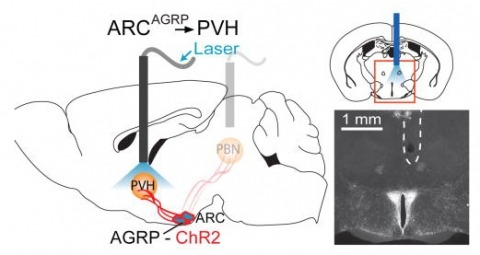
Hunger is a complex behavioural state that elicits intense food seeking and consumption. These behaviours are rapidly recapitulated by activation of starvation-sensitive AGRP neurons, which present an entry point for reverse-engineering neural circuits for hunger. Here we mapped synaptic interactions of AGRP neurons with multiple cell populations in mice and probed the contribution of these distinct circuits to feeding behaviour using optogenetic and pharmacogenetic techniques. An inhibitory circuit with paraventricular hypothalamus (PVH) neurons substantially accounted for acute AGRP neuron-evoked eating, whereas two other prominent circuits were insufficient. Within the PVH, we found that AGRP neurons target and inhibit oxytocin neurons, a small population that is selectively lost in Prader-Willi syndrome, a condition involving insatiable hunger. By developing strategies for evaluating molecularly defined circuits, we show that AGRP neuron suppression of oxytocin neurons is critical for evoked feeding. These experiments reveal a new neural circuit that regulates hunger state and pathways associated with overeating disorders.
The GABA transporters (GAT1, GAT2, GAT3, and BGT1) have mostly been discussed in relation to their potential roles in controlling the action of transmitter GABA in the nervous system. We have generated the first mice lacking the GAT2 (slc6a13) gene. Deletion of GAT2 (both mRNA and protein) neither affected growth, fertility, nor life span under nonchallenging rearing conditions. Immunocytochemistry showed that the GAT2 protein was predominantly expressed in the plasma membranes of periportal hepatocytes and in the basolateral membranes of proximal tubules in the renal cortex. This was validated by processing tissue from wild-type and knockout mice in parallel. Deletion of GAT2 reduced liver taurine levels by 50%, without affecting the expression of the taurine transporter TAUT. These results suggest an important role for GAT2 in taurine uptake from portal blood into liver. In support of this notion, GAT2-transfected HEK293 cells transported [(3)H]taurine. Furthermore, most of the uptake of [(3)H]GABA by cultured rat hepatocytes was due to GAT2, and this uptake was inhibited by taurine. GAT2 was not detected in brain parenchyma proper, excluding a role in GABA inactivation. It was, however, expressed in the leptomeninges and in a subpopulation of brain blood vessels. Deletion of GAT2 increased brain taurine levels by 20%, suggesting a taurine-exporting role for GAT2 in the brain.
Electronic and biological systems both perform complex information processing, but they use very different techniques. Though electronics has the advantage in raw speed, biological systems have the edge in many other areas. They can be produced, and indeed self-reproduce, without expensive and finicky factories. They are tolerant of manufacturing defects, and learn and adapt for better performance. In many cases they can self-repair damage. These advantages suggest that biological systems might be useful in a wide variety of tasks involving information processing. So far, all attempts to use the nervous system of a living organism for information processing have involved selective breeding of existing organisms. This approach, largely independent of the details of internal operation, is used since we do not yet understand how neural systems work, nor exactly how they are constructed. However, as our knowledge increases, the day will come when we can envision useful nervous systems and design them based upon what we want them to do, as opposed to variations on what has been already built. We will then need tools, corresponding to our Electronic Design Automation tools, to help with the design. This paper is concerned with what such tools might look like.
Somatic sexual dimorphisms outside of the nervous system in Drosophila melanogaster are largely controlled by the male- and female-specific Doublesex transcription factors (DSX(M) and DSX(F), respectively). The DSX proteins must act at the right times and places in development to regulate the diverse array of genes that sculpt male and female characteristics across a variety of tissues. To explore how cellular and developmental contexts integrate with doublesex (dsx) gene function, we focused on the sexually dimorphic number of gustatory sense organs (GSOs) in the foreleg. We show that DSX(M) and DSX(F) promote and repress GSO formation, respectively, and that their relative contribution to this dimorphism varies along the proximodistal axis of the foreleg. Our results suggest that the DSX proteins impact specification of the gustatory sensory organ precursors (SOPs). DSX(F) then acts later in the foreleg to regulate gustatory receptor neuron axon guidance. These results suggest that the foreleg provides a unique opportunity for examining the context-dependent functions of DSX.
Anatomy of large biological specimens is often reconstructed from serially sectioned volumes imaged by high-resolution microscopy. We developed a method to reassemble a continuous volume from such large section series that explicitly minimizes artificial deformation by applying a global elastic constraint. We demonstrate our method on a series of transmission electron microscopy sections covering the entire 558-cell Caenorhabditis elegans embryo and a segment of the Drosophila melanogaster larval ventral nerve cord.
Escape behaviors are, by necessity, fast and robust, making them excellent systems with which to study the neural basis of behavior. This is especially true in insects, which have comparatively tractable nervous systems and members who are amenable to manipulation with genetic tools. Recent technical developments in high-speed video reveal that, despite their short duration, insect escape behaviors are more complex than previously appreciated. For example, before initiating an escape jump, a fly performs sophisticated posture and stimulus-dependent preparatory leg movements that enable it to jump away from a looming threat. This newfound flexibility raises the question of how the nervous system generates a behavior that is both rapid and flexible. Recordings from the cricket nervous system suggest that synchrony between the activity of specific interneuron pairs may provide a rapid cue for the cricket to detect the direction of an approaching predator and thus which direction it should run. Technical advances make possible wireless recording from neurons while locusts escape from a looming threat, enabling, for the first time, a direct correlation between the activity of multiple neurons and the time-course of an insect escape behavior.
Two-photon probe excitation data are commonly presented as absorption cross section or molecular brightness (the detected fluorescence rate per molecule). We report two-photon molecular brightness spectra for a diverse set of organic and genetically encoded probes with an automated spectroscopic system based on fluorescence correlation spectroscopy. The two-photon action cross section can be extracted from molecular brightness measurements at low excitation intensities, while peak molecular brightness (the maximum molecular brightness with increasing excitation intensity) is measured at higher intensities at which probe photophysical effects become significant. The spectral shape of these two parameters was similar across all dye families tested. Peak molecular brightness spectra, which can be obtained rapidly and with reduced experimental complexity, can thus serve as a first-order approximation to cross-section spectra in determining optimal wavelengths for two-photon excitation, while providing additional information pertaining to probe photostability. The data shown should assist in probe choice and experimental design for multiphoton microscopy studies. Further, we show that, by the addition of a passive pulse splitter, nonlinear bleaching can be reduced-resulting in an enhancement of the fluorescence signal in fluorescence correlation spectroscopy by a factor of two. This increase in fluorescence signal, together with the observed resemblance of action cross section and peak brightness spectra, suggests higher-order photobleaching pathways for two-photon excitation.
Fluorescence imaging has revolutionized biomedical research over the past three decades. Its high molecular specificity and unrivalled single-molecule-level sensitivity have enabled breakthroughs in a number of research fields. For in vivo applications its major limitation is its superficial imaging depth, a result of random scattering in biological tissues causing exponential attenuation of the ballistic component of a light wave. Here, we present fluorescence imaging beyond the ballistic regime by combining single-cycle pulsed ultrasound modulation and digital optical phase conjugation. We demonstrate a near-isotropic three-dimensional localized sound–light interaction zone. With the exceptionally high optical gain provided by the digital optical phase conjugation system, we can deliver sufficient optical power to a focus inside highly scattering media for not only fluorescence imaging but also a variety of linear and nonlinear spectroscopy measurements. This technology paves the way for many important applications in both fundamental biology research and clinical studies.