Filter
Associated Lab
- Aguilera Castrejon Lab (1) Apply Aguilera Castrejon Lab filter
- Ahrens Lab (45) Apply Ahrens Lab filter
- Aso Lab (39) Apply Aso Lab filter
- Baker Lab (19) Apply Baker Lab filter
- Betzig Lab (98) Apply Betzig Lab filter
- Beyene Lab (4) Apply Beyene Lab filter
- Bock Lab (14) Apply Bock Lab filter
- Branson Lab (45) Apply Branson Lab filter
- Card Lab (34) Apply Card Lab filter
- Cardona Lab (44) Apply Cardona Lab filter
- Chklovskii Lab (10) Apply Chklovskii Lab filter
- Clapham Lab (11) Apply Clapham Lab filter
- Cui Lab (19) Apply Cui Lab filter
- Darshan Lab (8) Apply Darshan Lab filter
- Dickson Lab (32) Apply Dickson Lab filter
- Druckmann Lab (21) Apply Druckmann Lab filter
- Dudman Lab (34) Apply Dudman Lab filter
- Eddy/Rivas Lab (30) Apply Eddy/Rivas Lab filter
- Egnor Lab (4) Apply Egnor Lab filter
- Espinosa Medina Lab (12) Apply Espinosa Medina Lab filter
- Feliciano Lab (6) Apply Feliciano Lab filter
- Fetter Lab (31) Apply Fetter Lab filter
- Fitzgerald Lab (15) Apply Fitzgerald Lab filter
- Freeman Lab (15) Apply Freeman Lab filter
- Funke Lab (34) Apply Funke Lab filter
- Gonen Lab (59) Apply Gonen Lab filter
- Grigorieff Lab (34) Apply Grigorieff Lab filter
- Harris Lab (48) Apply Harris Lab filter
- Heberlein Lab (13) Apply Heberlein Lab filter
- Hermundstad Lab (17) Apply Hermundstad Lab filter
- Hess Lab (68) Apply Hess Lab filter
- Ilanges Lab (1) Apply Ilanges Lab filter
- Jayaraman Lab (39) Apply Jayaraman Lab filter
- Ji Lab (33) Apply Ji Lab filter
- Johnson Lab (1) Apply Johnson Lab filter
- Karpova Lab (13) Apply Karpova Lab filter
- Keleman Lab (8) Apply Keleman Lab filter
- Keller Lab (60) Apply Keller Lab filter
- Lavis Lab (123) Apply Lavis Lab filter
- Lee (Albert) Lab (29) Apply Lee (Albert) Lab filter
- Leonardo Lab (19) Apply Leonardo Lab filter
- Li Lab (1) Apply Li Lab filter
- Lippincott-Schwartz Lab (89) Apply Lippincott-Schwartz Lab filter
- Liu (Zhe) Lab (53) Apply Liu (Zhe) Lab filter
- Looger Lab (136) Apply Looger Lab filter
- Magee Lab (31) Apply Magee Lab filter
- Menon Lab (12) Apply Menon Lab filter
- Murphy Lab (6) Apply Murphy Lab filter
- O'Shea Lab (3) Apply O'Shea Lab filter
- Otopalik Lab (1) Apply Otopalik Lab filter
- Pachitariu Lab (28) Apply Pachitariu Lab filter
- Pastalkova Lab (5) Apply Pastalkova Lab filter
- Pavlopoulos Lab (7) Apply Pavlopoulos Lab filter
- Pedram Lab (3) Apply Pedram Lab filter
- Podgorski Lab (16) Apply Podgorski Lab filter
- Reiser Lab (43) Apply Reiser Lab filter
- Riddiford Lab (20) Apply Riddiford Lab filter
- Romani Lab (28) Apply Romani Lab filter
- Rubin Lab (101) Apply Rubin Lab filter
- Saalfeld Lab (43) Apply Saalfeld Lab filter
- Satou Lab (1) Apply Satou Lab filter
- Scheffer Lab (36) Apply Scheffer Lab filter
- Schreiter Lab (44) Apply Schreiter Lab filter
- Shroff Lab (22) Apply Shroff Lab filter
- Simpson Lab (18) Apply Simpson Lab filter
- Singer Lab (37) Apply Singer Lab filter
- Spruston Lab (55) Apply Spruston Lab filter
- Stern Lab (69) Apply Stern Lab filter
- Sternson Lab (47) Apply Sternson Lab filter
- Stringer Lab (25) Apply Stringer Lab filter
- Svoboda Lab (131) Apply Svoboda Lab filter
- Tebo Lab (7) Apply Tebo Lab filter
- Tervo Lab (9) Apply Tervo Lab filter
- Tillberg Lab (14) Apply Tillberg Lab filter
- Tjian Lab (17) Apply Tjian Lab filter
- Truman Lab (58) Apply Truman Lab filter
- Turaga Lab (34) Apply Turaga Lab filter
- Turner Lab (24) Apply Turner Lab filter
- Vale Lab (6) Apply Vale Lab filter
- Voigts Lab (1) Apply Voigts Lab filter
- Wang (Meng) Lab (9) Apply Wang (Meng) Lab filter
- Wang (Shaohe) Lab (5) Apply Wang (Shaohe) Lab filter
- Wu Lab (8) Apply Wu Lab filter
- Zlatic Lab (26) Apply Zlatic Lab filter
- Zuker Lab (5) Apply Zuker Lab filter
Associated Project Team
- CellMap (4) Apply CellMap filter
- COSEM (3) Apply COSEM filter
- Fly Descending Interneuron (10) Apply Fly Descending Interneuron filter
- Fly Functional Connectome (14) Apply Fly Functional Connectome filter
- Fly Olympiad (5) Apply Fly Olympiad filter
- FlyEM (51) Apply FlyEM filter
- FlyLight (46) Apply FlyLight filter
- GENIE (40) Apply GENIE filter
- Integrative Imaging (1) Apply Integrative Imaging filter
- Larval Olympiad (2) Apply Larval Olympiad filter
- MouseLight (16) Apply MouseLight filter
- NeuroSeq (1) Apply NeuroSeq filter
- ThalamoSeq (1) Apply ThalamoSeq filter
- Tool Translation Team (T3) (24) Apply Tool Translation Team (T3) filter
- Transcription Imaging (45) Apply Transcription Imaging filter
Associated Support Team
- Project Pipeline Support (1) Apply Project Pipeline Support filter
- Anatomy and Histology (18) Apply Anatomy and Histology filter
- Cryo-Electron Microscopy (33) Apply Cryo-Electron Microscopy filter
- Electron Microscopy (12) Apply Electron Microscopy filter
- Fly Facility (39) Apply Fly Facility filter
- Gene Targeting and Transgenics (11) Apply Gene Targeting and Transgenics filter
- Integrative Imaging (10) Apply Integrative Imaging filter
- Janelia Experimental Technology (35) Apply Janelia Experimental Technology filter
- Management Team (1) Apply Management Team filter
- Molecular Genomics (15) Apply Molecular Genomics filter
- Primary & iPS Cell Culture (13) Apply Primary & iPS Cell Culture filter
- Project Technical Resources (36) Apply Project Technical Resources filter
- Quantitative Genomics (19) Apply Quantitative Genomics filter
- Scientific Computing Software (59) Apply Scientific Computing Software filter
- Scientific Computing Systems (6) Apply Scientific Computing Systems filter
- Viral Tools (14) Apply Viral Tools filter
- Vivarium (6) Apply Vivarium filter
Publication Date
- 2024 (132) Apply 2024 filter
- 2023 (175) Apply 2023 filter
- 2022 (166) Apply 2022 filter
- 2021 (174) Apply 2021 filter
- 2020 (177) Apply 2020 filter
- 2019 (177) Apply 2019 filter
- 2018 (206) Apply 2018 filter
- 2017 (186) Apply 2017 filter
- 2016 (191) Apply 2016 filter
- 2015 (195) Apply 2015 filter
- 2014 (190) Apply 2014 filter
- 2013 (136) Apply 2013 filter
- 2012 (112) Apply 2012 filter
- 2011 (98) Apply 2011 filter
- 2010 (61) Apply 2010 filter
- 2009 (56) Apply 2009 filter
- 2008 (40) Apply 2008 filter
- 2007 (21) Apply 2007 filter
- 2006 (3) Apply 2006 filter
2496 Janelia Publications
Showing 2451-2460 of 2496 results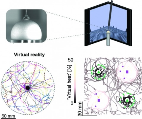
Studying the intertwined roles of sensation, experience, and directed action in navigation has been facilitated by the development of virtual reality (VR) environments for head-fixed animals, allowing for quantitative measurements of behavior in well-controlled conditions. VR has long featured in studies of Drosophila melanogaster, but these experiments have typically allowed the fly to change only its heading in a visual scene and not its position. Here we explore how flies move in two dimensions (2D) using a visual VR environment that more closely captures an animal's experience during free behavior. We show that flies' 2D interaction with landmarks cannot be automatically derived from their orienting behavior under simpler one-dimensional (1D) conditions. Using novel paradigms, we then demonstrate that flies in 2D VR adapt their behavior in response to optogenetically delivered appetitive and aversive stimuli. Much like free-walking flies after encounters with food, head-fixed flies exploring a 2D VR respond to optogenetic activation of sugar-sensing neurons by initiating a local search, which appears not to rely on visual landmarks. Visual landmarks can, however, help flies to avoid areas in VR where they experience an aversive, optogenetically generated heat stimulus. By coupling aversive virtual heat to the flies' presence near visual landmarks of specific shapes, we elicit selective learned avoidance of those landmarks. Thus, we demonstrate that head-fixed flies adaptively navigate in 2D virtual environments, but their reliance on visual landmarks is context dependent. These behavioral paradigms set the stage for interrogation of the fly brain circuitry underlying flexible navigation in complex multisensory environments.
The hippocampus is critical for recollecting and imagining experiences. This is believed to involve voluntarily drawing from hippocampal memory representations of people, events, and places, including maplike representations of familiar environments. However, whether representations in such "cognitive maps" can be volitionally accessed is unknown. We developed a brain-machine interface to test whether rats can do so by controlling their hippocampal activity in a flexible, goal-directed, and model-based manner. We found that rats can efficiently navigate or direct objects to arbitrary goal locations within a virtual reality arena solely by activating and sustaining appropriate hippocampal representations of remote places. This provides insight into the mechanisms underlying episodic memory recall, mental simulation and planning, and imagination and opens up possibilities for high-level neural prosthetics that use hippocampal representations.
Voltage imaging enables monitoring neural activity at sub-millisecond and sub-cellular scale, unlocking the study of subthreshold activity, synchrony, and network dynamics with unprecedented spatio-temporal resolution. However, high data rates (>800MB/s) and low signal-to-noise ratios create bottlenecks for analyzing such datasets. Here we present VolPy, an automated and scalable pipeline to pre-process voltage imaging datasets. VolPy features motion correction, memory mapping, automated segmentation, denoising and spike extraction, all built on a highly parallelizable, modular, and extensible framework optimized for memory and speed. To aid automated segmentation, we introduce a corpus of 24 manually annotated datasets from different preparations, brain areas and voltage indicators. We benchmark VolPy against ground truth segmentation, simulations and electrophysiology recordings, and we compare its performance with existing algorithms in detecting spikes. Our results indicate that VolPy's performance in spike extraction and scalability are state-of-the-art.
Neurons integrate synaptic inputs within their dendrites and produce spiking outputs, which then propagate down the axon and back into the dendrites where they contribute to plasticity. Mapping the voltage dynamics in dendritic arbors of live animals is crucial for understanding neuronal computation and plasticity rules. Here we combine patterned channelrhodopsin activation with dual-plane structured illumination voltage imaging, for simultaneous perturbation and monitoring of dendritic and somatic voltage in Layer 2/3 pyramidal neurons in anesthetized and awake mice. We examined the integration of synaptic inputs and compared the dynamics of optogenetically evoked, spontaneous, and sensory-evoked back-propagating action potentials (bAPs). Our measurements revealed a broadly shared membrane voltage throughout the dendritic arbor, and few signatures of electrical compartmentalization among synaptic inputs. However, we observed spike rate acceleration-dependent propagation of bAPs into distal dendrites. We propose that this dendritic filtering of bAPs may play a critical role in activity-dependent plasticity.
Motor systems must continuously adapt their output to maintain a desired trajectory. While the spinal circuits underlying rhythmic locomotion are well described, little is known about how the network modulates its output strength. A major challenge has been the difficulty of recording from spinal neurons during behavior. Here, we use voltage imaging to map the membrane potential of large populations of glutamatergic neurons throughout the spinal cord of the larval zebrafish during fictive swimming in a virtual environment. We characterized a previously undescribed subpopulation of tonic-spiking ventral V3 neurons whose spike rate correlated with swimming strength and bout length. Optogenetic activation of V3 neurons led to stronger swimming and longer bouts but did not affect tail beat frequency. Genetic ablation of V3 neurons led to reduced locomotor adaptation. The power of voltage imaging allowed us to identify V3 neurons as a critical driver of locomotor adaptation in zebrafish.
Motor systems must continuously adapt their output to maintain a desired trajectory. While the spinal circuits underlying rhythmic locomotion are well described, little is known about how the network modulates its output strength. A major challenge has been the difficulty of recording from spinal neurons during behavior. Here, we use voltage imaging to map the membrane potential of large populations of glutamatergic neurons throughout the spinal cord of the larval zebrafish during fictive swimming in a virtual environment. We characterized a previously undescribed subpopulation of tonic-spiking ventral V3 neurons whose spike rate correlated with swimming strength and bout length. Optogenetic activation of V3 neurons led to stronger swimming and longer bouts but did not affect tail beat frequency. Genetic ablation of V3 neurons led to reduced locomotor adaptation. The power of voltage imaging allowed us to identify V3 neurons as a critical driver of locomotor adaptation in zebrafish.
Voltage-gated ion channels support electrochemical activity in cells and are largely responsible for information flow throughout the nervous systems. The voltage sensor domains in these channels sense changes in transmembrane potential and control ion flux across membranes. The X-ray structures of a few voltage-gated ion channels in detergents have been determined and have revealed clear structural variations among their respective voltage sensor domains. More recent studies demonstrated that lipids around a voltage-gated channel could directly alter its conformational state in membrane. Because of these disparities, the structural basis for voltage sensing in native membranes remains elusive. Here, through electron-crystallographic analysis of membrane-embedded proteins, we present the detailed view of a voltage-gated potassium channel in its inactivated state. Contrary to all known structures of voltage-gated ion channels in detergents, our data revealed a unique conformation in which the four voltage sensor domains of a voltage-gated potassium channel from Aeropyrum pernix (KvAP) form a ring structure that completely surrounds the pore domain of the channel. Such a structure is named the voltage sensor ring. Our biochemical and electrophysiological studies support that the voltage sensor ring represents a physiological conformation. These data together suggest that lipids exert strong effects on the channel structure and that these effects may be changed upon membrane disruption. Our results have wide implications for lipid-protein interactions in general and for the mechanism of voltage sensing in particular.
In rats, navigating through an environment requires continuous information about objects near the head. Sensory information such as object location and surface texture are encoded by spike firing patterns of single neurons within rat barrel cortex. Although there are many studies using single-unit electrophysiology, much less is known regarding the spatiotemporal pattern of activity of populations of neurons in barrel cortex in response to whisker stimulation. To examine cortical response at the population level, we used voltage-sensitive dye (VSD) imaging to examine ensemble spatiotemporal dynamics of barrel cortex in response to stimulation of single or two adjacent whiskers in urethane-anesthetized rats. Single whisker stimulation produced a poststimulus fluorescence response peak within 12-16 ms in the barrel corresponding to the stimulated whisker (principal whisker). This fluorescence subsequently propagated throughout the barrel field, spreading anisotropically preferentially along a barrel row. After paired whisker stimulation, the VSD signal showed sublinear summation (less than the sum of 2 single whisker stimulations), consistent with previous electrophysiological and imaging studies. Surprisingly, we observed a spatial shift in the center of activation occurring over a 10- to 20-ms period with shift magnitudes of 1-2 barrels. This shift occurred predominantly in the posteromedial direction within the barrel field. Our data thus reveal previously unreported spatiotemporal patterns of barrel cortex activation. We suggest that this nontopographical shift is consistent with known functional and anatomic asymmetries in barrel cortex and that it may provide an important insight for understanding barrel field activation during whisking behavior.
The last decade has seen a rapid increase in the number of tools to acquire volume electron microscopy (EM) data. Several new scanning EM (SEM) imaging methods have emerged, and classical transmission EM (TEM) methods are being scaled up and automated. Here we summarize the new methods for acquiring large EM volumes, and discuss the tradeoffs in terms of resolution, acquisition speed, and reliability. We then assess each method’s applicability to the problem of reconstructing anatomical connectivity between neurons, considering both the current capabilities and future prospects of the method. Finally, we argue that neuronal ’wiring diagrams’ are likely necessary, but not sufficient, to understand the operation of most neuronal circuits: volume EM imaging will likely find its best application in combination with other methods in neuroscience, such as molecular biology, optogenetics, and physiology.
Life exists in three dimensions, but until the turn of the century most electron microscopy methods provided only 2D image data. Recently, electron microscopy techniques capable of delving deep into the structure of cells and tissues have emerged, collectively called volume electron microscopy (vEM). Developments in vEM have been dubbed a quiet revolution as the field evolved from established transmission and scanning electron microscopy techniques, so early publications largely focused on the bioscience applications rather than the underlying technological breakthroughs. However, with an explosion in the uptake of vEM across the biosciences and fast-paced advances in volume, resolution, throughput and ease of use, it is timely to introduce the field to new audiences. In this Primer, we introduce the different vEM imaging modalities, the specialized sample processing and image analysis pipelines that accompany each modality and the types of information revealed in the data. We showcase key applications in the biosciences where vEM has helped make breakthrough discoveries and consider limitations and future directions. We aim to show new users how vEM can support discovery science in their own research fields and inspire broader uptake of the technology, finally allowing its full adoption into mainstream biological imaging.