Filter
Associated Lab
- Aso Lab (1) Apply Aso Lab filter
- Baker Lab (3) Apply Baker Lab filter
- Betzig Lab (7) Apply Betzig Lab filter
- Bock Lab (1) Apply Bock Lab filter
- Cui Lab (3) Apply Cui Lab filter
- Dickson Lab (1) Apply Dickson Lab filter
- Dudman Lab (1) Apply Dudman Lab filter
- Eddy/Rivas Lab (5) Apply Eddy/Rivas Lab filter
- Fetter Lab (2) Apply Fetter Lab filter
- Gonen Lab (1) Apply Gonen Lab filter
- Hess Lab (2) Apply Hess Lab filter
- Jayaraman Lab (2) Apply Jayaraman Lab filter
- Ji Lab (2) Apply Ji Lab filter
- Keller Lab (2) Apply Keller Lab filter
- Lavis Lab (5) Apply Lavis Lab filter
- Lee (Albert) Lab (1) Apply Lee (Albert) Lab filter
- Leonardo Lab (2) Apply Leonardo Lab filter
- Looger Lab (7) Apply Looger Lab filter
- Magee Lab (1) Apply Magee Lab filter
- Menon Lab (3) Apply Menon Lab filter
- Murphy Lab (1) Apply Murphy Lab filter
- Reiser Lab (2) Apply Reiser Lab filter
- Riddiford Lab (1) Apply Riddiford Lab filter
- Rubin Lab (3) Apply Rubin Lab filter
- Scheffer Lab (2) Apply Scheffer Lab filter
- Schreiter Lab (2) Apply Schreiter Lab filter
- Simpson Lab (3) Apply Simpson Lab filter
- Singer Lab (1) Apply Singer Lab filter
- Sternson Lab (6) Apply Sternson Lab filter
- Svoboda Lab (7) Apply Svoboda Lab filter
- Tjian Lab (2) Apply Tjian Lab filter
- Truman Lab (1) Apply Truman Lab filter
- Zlatic Lab (1) Apply Zlatic Lab filter
- Zuker Lab (2) Apply Zuker Lab filter
Associated Project Team
Publication Date
- December 2011 (10) Apply December 2011 filter
- November 2011 (8) Apply November 2011 filter
- October 2011 (8) Apply October 2011 filter
- September 2011 (8) Apply September 2011 filter
- August 2011 (9) Apply August 2011 filter
- July 2011 (5) Apply July 2011 filter
- June 2011 (10) Apply June 2011 filter
- May 2011 (6) Apply May 2011 filter
- April 2011 (5) Apply April 2011 filter
- March 2011 (6) Apply March 2011 filter
- February 2011 (8) Apply February 2011 filter
- January 2011 (15) Apply January 2011 filter
- Remove 2011 filter 2011
98 Janelia Publications
Showing 51-60 of 98 resultsThe mammalian brain is best understood as a multi-scale hierarchical neural system, in the sense that connection and function occur on multiple scales from micro to macro. Modern genomic-scale expression profiling can provide insight into methodologies that elucidate this architecture. We present a methodology for understanding the relationship of gene expression and neuroanatomy based on correlation between gene expression profiles across tissue samples. A resulting tool, NeuroBlast, can identify networks of genes co-expressed within or across neuroanatomic structures. The method applies to any data modality that can be mapped with sufficient spatial resolution, and provides a computation technique to elucidate neuroanatomy via patterns of gene expression on spatial and temporal scales. In addition, from the perspective of spatial location, we discuss a complementary technique that identifies gene classes that contribute to defining anatomic patterns.
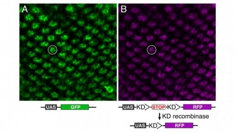
Site-specific recombinases have been used for two decades to manipulate the structure of animal genomes in highly predictable ways and have become major research tools. However, the small number of recombinases demonstrated to have distinct specificities, low toxicity, and sufficient activity to drive reactions to completion in animals has been a limitation. In this report we show that four recombinases derived from yeast-KD, B2, B3, and R-are highly active and nontoxic in Drosophila and that KD, B2, B3, and the widely used FLP recombinase have distinct target specificities. We also show that the KD and B3 recombinases are active in mice.
Aversive olfactory memory is formed in the mushroom bodies in Drosophila melanogaster. Memory retrieval requires mushroom body output, but the manner in which a memory trace in the mushroom body drives conditioned avoidance of a learned odor remains unknown. To identify neurons that are involved in olfactory memory retrieval, we performed an anatomical and functional screen of defined sets of mushroom body output neurons. We found that MB-V2 neurons were essential for retrieval of both short- and long-lasting memory, but not for memory formation or memory consolidation. MB-V2 neurons are cholinergic efferent neurons that project from the mushroom body vertical lobes to the middle superiormedial protocerebrum and the lateral horn. Notably, the odor response of MB-V2 neurons was modified after conditioning. As the lateral horn has been implicated in innate responses to repellent odorants, we propose that MB-V2 neurons recruit the olfactory pathway involved in innate odor avoidance during memory retrieval.

When the contrast of an image flickers as it moves, humans perceive an illusory reversal in the direction of motion. This classic illusion, called reverse-phi motion, has been well-characterized using psychophysics, and several models have been proposed to account for its effects. Here, we show that Drosophila melanogaster also respond behaviorally to the reverse-phi illusion and that the illusion is present in dendritic calcium signals of motion-sensitive neurons in the fly lobula plate. These results closely match the predictions of the predominant model of fly motion detection. However, high flicker rates cause an inversion of the reverse-phi behavioral response that is also present in calcium signals of lobula plate tangential cell dendrites but not predicted by the model. The fly’s behavioral and neural responses to the reverse-phi illusion reveal unexpected interactions between motion and flicker signals in the fly visual system and suggest that a similar correlation-based mechanism underlies visual motion detection across the animal kingdom.
Because of its genetic, molecular, and behavioral tractability, Drosophila has emerged as a powerful model system for studying molecular and cellular mechanisms underlying the development and function of nervous systems. The Drosophila nervous system has fewer neurons and exhibits a lower glia:neuron ratio than is seen in vertebrate nervous systems. Despite the simplicity of the Drosophila nervous system, glial organization in flies is as sophisticated as it is in vertebrates. Furthermore, fly glial cells play vital roles in neural development and behavior. In addition, powerful genetic tools are continuously being created to explore cell function in vivo. In taking advantage of these features, the fly nervous system serves as an excellent model system to study general aspects of glial cell development and function in vivo. In this article, we review and discuss advanced genetic tools that are potentially useful for understanding glial cell biology in Drosophila.
In this issue of Neuron, Makino and Malinow and Kleindienst et al. present evidence of a behaviorally induced form of synaptic plasticity that would encourage the development of fine-scale structured input patterns and the binding of features within single neurons.
Research on the biology of addiction has advanced significantly over the last 50 years expanding our understanding of the brain mechanisms underlying reward, reinforcement and craving. Novel experimental approaches and techniques have provided an ever increasing armory of tools to dissect behavioral processes, neural networks and molecular mechanisms. The ultimate goal is to reintegrate this knowledge into a coherent, mechanistic framework of addiction to help identify new treatment. This can be greatly facilitated by using tools that allow, with great spatial and temporal specificity, to link molecular changes with altered activation of neural circuits and behavior. Such specificity can now be achieved by using optogenetic tools. Our review describes the general principles of optogenetics and its use to understand the links between neural activity and behavior. We also provide an overview of recent studies using optogenetic tools in addiction and consider some outstanding questions of addiction research that are particularly amenable for optogenetic approaches.
News & Views | Published: 27 December 2010 Nature Neuroscience volume 14, pages 6–7 (2011) | Download Citation Pruning of excess branches is essential for the maturation of developing neuronal circuits. Cross-talk between TGF-β signaling and two antagonistic orphan nuclear receptors governs the pruning of larval γ neurons in the Drosophila pupa. Neural circuits are remodeled as the brain matures or acquires new functions. Such developmental remodeling involves complex cellular changes that are tightly regulated in space and time. During metamorphosis of holometabolous insect brains, most larval functional neurons are rewired into the adult circuitry, and study of these processes has been particularly fruitful for the elucidation of the mechanisms that underlie neuron remodeling1. In metamorphosing Drosophila, nuclear signaling of the steroid hormone receptor ecdysone receptor B1 isoform (EcR-B1) cell-autonomously orchestrates neuron remodeling. Only neurons destined to remodel upregulate EcR-B1 expression before a crucial pre-pupal ecdysone pulse2. It is therefore necessary to determine the mechanisms that pattern EcR-B1 expression to understand how developmental neuronal remodeling is programmed in Drosophila.Orphan nuclear receptors control neuronal remodeling during fly metamorphosis
Live-cell fluorescence light microscopy has emerged as an important tool in the study of cellular biology. The development of fluorescent markers in parallel with super-resolution imaging systems has pushed light microscopy into the realm of molecular visualization at the nanometer scale. Resolutions previously only attained with electron microscopes are now within the grasp of light microscopes. However, until recently, live-cell imaging approaches have eluded super-resolution microscopy, hampering it from reaching its full potential for revealing the dynamic interactions in biology occurring at the single molecule level. Here we examine recent advances in the super-resolution imaging of living cells by reviewing recent breakthroughs in single molecule localization microscopy methods such as PALM and STORM to achieve this important goal.
A parallel wavefront optimization method is demonstrated experimentally to focus light through random scattering media. The simultaneous modulation of multiple phase elements, each at a unique frequency, enables a parallel determination of the optimal wavefront. Compared to a pixel-by-pixel measurement, the reported parallel method uses the target signal in a highly efficient way. With 441 phase elements, a high-quality focus was formed through a glass diffuser with a peak-to-background ratio of \~{}270. The accuracy and repeatability of the system were tested through experiments.