Filter
Associated Lab
- Ahrens Lab (7) Apply Ahrens Lab filter
- Aso Lab (2) Apply Aso Lab filter
- Baker Lab (2) Apply Baker Lab filter
- Betzig Lab (12) Apply Betzig Lab filter
- Bock Lab (1) Apply Bock Lab filter
- Branson Lab (5) Apply Branson Lab filter
- Card Lab (3) Apply Card Lab filter
- Cardona Lab (8) Apply Cardona Lab filter
- Dickson Lab (4) Apply Dickson Lab filter
- Druckmann Lab (3) Apply Druckmann Lab filter
- Dudman Lab (3) Apply Dudman Lab filter
- Eddy/Rivas Lab (1) Apply Eddy/Rivas Lab filter
- Egnor Lab (1) Apply Egnor Lab filter
- Fetter Lab (6) Apply Fetter Lab filter
- Fitzgerald Lab (1) Apply Fitzgerald Lab filter
- Freeman Lab (4) Apply Freeman Lab filter
- Funke Lab (2) Apply Funke Lab filter
- Gonen Lab (8) Apply Gonen Lab filter
- Grigorieff Lab (6) Apply Grigorieff Lab filter
- Harris Lab (5) Apply Harris Lab filter
- Hess Lab (2) Apply Hess Lab filter
- Jayaraman Lab (3) Apply Jayaraman Lab filter
- Ji Lab (4) Apply Ji Lab filter
- Kainmueller Lab (3) Apply Kainmueller Lab filter
- Karpova Lab (3) Apply Karpova Lab filter
- Keleman Lab (1) Apply Keleman Lab filter
- Keller Lab (6) Apply Keller Lab filter
- Lavis Lab (13) Apply Lavis Lab filter
- Lee (Albert) Lab (1) Apply Lee (Albert) Lab filter
- Leonardo Lab (2) Apply Leonardo Lab filter
- Li Lab (2) Apply Li Lab filter
- Lippincott-Schwartz Lab (9) Apply Lippincott-Schwartz Lab filter
- Liu (Zhe) Lab (4) Apply Liu (Zhe) Lab filter
- Looger Lab (10) Apply Looger Lab filter
- Magee Lab (2) Apply Magee Lab filter
- Menon Lab (2) Apply Menon Lab filter
- Murphy Lab (2) Apply Murphy Lab filter
- Pachitariu Lab (2) Apply Pachitariu Lab filter
- Pastalkova Lab (1) Apply Pastalkova Lab filter
- Pavlopoulos Lab (3) Apply Pavlopoulos Lab filter
- Pedram Lab (1) Apply Pedram Lab filter
- Podgorski Lab (1) Apply Podgorski Lab filter
- Reiser Lab (3) Apply Reiser Lab filter
- Romani Lab (2) Apply Romani Lab filter
- Rubin Lab (3) Apply Rubin Lab filter
- Saalfeld Lab (3) Apply Saalfeld Lab filter
- Schreiter Lab (2) Apply Schreiter Lab filter
- Simpson Lab (1) Apply Simpson Lab filter
- Singer Lab (5) Apply Singer Lab filter
- Spruston Lab (6) Apply Spruston Lab filter
- Stern Lab (7) Apply Stern Lab filter
- Sternson Lab (3) Apply Sternson Lab filter
- Stringer Lab (1) Apply Stringer Lab filter
- Svoboda Lab (8) Apply Svoboda Lab filter
- Tervo Lab (2) Apply Tervo Lab filter
- Tillberg Lab (1) Apply Tillberg Lab filter
- Tjian Lab (3) Apply Tjian Lab filter
- Truman Lab (11) Apply Truman Lab filter
- Turaga Lab (2) Apply Turaga Lab filter
- Turner Lab (1) Apply Turner Lab filter
- Wang (Shaohe) Lab (2) Apply Wang (Shaohe) Lab filter
- Wu Lab (1) Apply Wu Lab filter
- Zlatic Lab (2) Apply Zlatic Lab filter
Associated Project Team
- Fly Functional Connectome (2) Apply Fly Functional Connectome filter
- Fly Olympiad (1) Apply Fly Olympiad filter
- FlyEM (1) Apply FlyEM filter
- GENIE (2) Apply GENIE filter
- MouseLight (2) Apply MouseLight filter
- Tool Translation Team (T3) (1) Apply Tool Translation Team (T3) filter
- Transcription Imaging (11) Apply Transcription Imaging filter
Publication Date
- December 2016 (18) Apply December 2016 filter
- November 2016 (15) Apply November 2016 filter
- October 2016 (22) Apply October 2016 filter
- September 2016 (11) Apply September 2016 filter
- August 2016 (13) Apply August 2016 filter
- July 2016 (15) Apply July 2016 filter
- June 2016 (25) Apply June 2016 filter
- May 2016 (23) Apply May 2016 filter
- April 2016 (14) Apply April 2016 filter
- March 2016 (15) Apply March 2016 filter
- February 2016 (23) Apply February 2016 filter
- January 2016 (15) Apply January 2016 filter
- Remove 2016 filter 2016
Type of Publication
209 Publications
Showing 21-30 of 209 resultsThe physiology of N-methyl-d-aspartate (NMDA) receptors is fundamental to brain development and function. NMDA receptors are ionotropic glutamate receptors that function as heterotetramers composed mainly of GluN1 and GluN2 subunits. Activation of NMDA receptors requires binding of neurotransmitter agonists to a ligand-binding domain (LBD) and structural rearrangement of an amino-terminal domain (ATD). Recent crystal structures of GluN1-GluN2B NMDA receptors bound to agonists and an allosteric inhibitor, ifenprodil, represent the allosterically inhibited state. However, how the ATD and LBD move to activate the NMDA receptor ion channel remains unclear. Here we applied X-ray crystallography, single-particle electron cryomicroscopy and electrophysiology to rat NMDA receptors to show that, in the absence of ifenprodil, the bi-lobed structure of GluN2 ATD adopts an open conformation accompanied by rearrangement of the GluN1-GluN2 ATD heterodimeric interface, altering subunit orientation in the ATD and LBD and forming an active receptor conformation that gates the ion channel.
UNLABELLED: Neurons respond to specific features of sensory stimuli. In the visual system, for example, some neurons respond to motion of small but not large objects, whereas other neurons prefer motion of the entire visual field. Separate neurons respond equally to local and global motion but selectively to additional features of visual stimuli. How and where does response selectivity emerge? Here, we show that wide-field (WF) cells in retino-recipient layers of the mouse superior colliculus (SC) respond selectively to small moving objects. Moreover, we identify two mechanisms that contribute to this selectivity. First, we show that input restricted to a small portion of the broad dendritic arbor of WF cells is sufficient to trigger dendritic spikes that reliably propagate to the soma/axon. In vivo whole-cell recordings reveal that nearly every action potential evoked by visual stimuli has characteristics of spikes initiated in dendrites. Second, inhibitory input from a different class of SC neuron, horizontal cells, constrains the range of stimuli to which WF cells respond. Horizontal cells respond preferentially to the sudden appearance or rapid movement of large stimuli. Optogenetic reduction of their activity reduces movement selectivity and broadens size tuning in WF cells by increasing the relative strength of responses to stimuli that appear suddenly or cover a large region of space. Therefore, strongly propagating dendritic spikes enable small stimuli to drive spike output in WF cells and local inhibition helps restrict responses to stimuli that are both small and moving. SIGNIFICANCE STATEMENT: How do neurons respond selectively to some sensory stimuli but not others? In the visual system, a particularly relevant stimulus feature is object motion, which often reveals other animals. Here, we show how specific cells in the superior colliculus, one synapse downstream of the retina, respond selectively to object motion. These wide-field (WF) cells respond strongly to small objects that move slowly anywhere through a large region of space, but not to stationary objects or full-field motion. Action potential initiation in dendrites enables small stimuli to trigger visual responses and inhibitory input from cells that prefer large, suddenly appearing, or quickly moving stimuli restricts responses of WF cells to objects that are small and moving.
GAL4 gene expression imaging using confocal microscopy is a common and powerful technique used to study the nervous system of a model organism such as Drosophila melanogaster. Recent research projects focused on high throughput screenings of thousands of different driver lines, resulting in large image databases. The amount of data generated makes manual assessment tedious or even impossible. The first and most important step in any automatic image processing and data extraction pipeline is to enhance areas with relevant signal. However, data acquired via high throughput imaging tends to be less then ideal for this task, often showing high amounts of background signal. Furthermore, neuronal structures and in particular thin and elongated projections with a weak staining signal are easily lost. In this paper we present a method for enhancing the relevant signal by utilizing a Hessian-based filter to augment thin and weak tube-like structures in the image. To get optimal results, we present a novel adaptive background-aware enhancement filter parametrized with the local background intensity, which is estimated based on a common background model. We also integrate recent research on adaptive image enhancement into our approach, allowing us to propose an effective solution for known problems present in confocal microscopy images. We provide an evaluation based on annotated image data and compare our results against current state-of-the-art algorithms. The results show that our algorithm clearly outperforms the existing solutions.
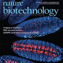
Optimal image quality in light-sheet microscopy requires a perfect overlap between the illuminating light sheet and the focal plane of the detection objective. However, mismatches between the light-sheet and detection planes are common owing to the spatiotemporally varying optical properties of living specimens. Here we present the AutoPilot framework, an automated method for spatiotemporally adaptive imaging that integrates (i) a multi-view light-sheet microscope capable of digitally translating and rotating light-sheet and detection planes in three dimensions and (ii) a computational method that continuously optimizes spatial resolution across the specimen volume in real time. We demonstrate long-term adaptive imaging of entire developing zebrafish (Danio rerio) and Drosophila melanogaster embryos and perform adaptive whole-brain functional imaging in larval zebrafish. Our method improves spatial resolution and signal strength two to five-fold, recovers cellular and sub-cellular structures in many regions that are not resolved by non-adaptive imaging, adapts to spatiotemporal dynamics of genetically encoded fluorescent markers and robustly optimizes imaging performance during large-scale morphogenetic changes in living organisms.
Nervous systems are composed of various cell types, but the extent of cell type diversity is poorly understood. We constructed a cellular taxonomy of one cortical region, primary visual cortex, in adult mice on the basis of single-cell RNA sequencing. We identified 49 transcriptomic cell types, including 23 GABAergic, 19 glutamatergic and 7 non-neuronal types. We also analyzed cell type-specific mRNA processing and characterized genetic access to these transcriptomic types by many transgenic Cre lines. Finally, we found that some of our transcriptomic cell types displayed specific and differential electrophysiological and axon projection properties, thereby confirming that the single-cell transcriptomic signatures can be associated with specific cellular properties.
Mitochondrial damage is the major factor underlying drug-induced liver disease but whether conditions that thwart mitochondrial injury can prevent or reverse drug-induced liver damage is unclear. A key molecule regulating mitochondria quality control is AMP activated kinase (AMPK). When activated, AMPK causes mitochondria to elongate/fuse and proliferate, with mitochondria now producing more ATP and less reactive oxygen species. Autophagy is also triggered, a process capable of removing damaged/defective mitochondria. To explore whether AMPK activation could potentially prevent or reverse the effects of drug-induced mitochondrial and hepatocellular damage, we added an AMPK activator to collagen sandwich cultures of rat and human hepatocytes exposed to the hepatotoxic drugs, acetaminophen or diclofenac. In the absence of AMPK activation, the drugs caused hepatocytes to lose polarized morphology and have significantly decreased ATP levels and viability. At the subcellular level, mitochondria underwent fragmentation and had decreased membrane potential due to decreased expression of the mitochondrial fusion proteins Mfn1, 2 and/or Opa1. Adding AICAR, a specific AMPK activator, at the time of drug exposure prevented and reversed these effects. The mitochondria became highly fused and ATP production increased, and hepatocytes maintained polarized morphology. In exploring the mechanism responsible for this preventive and reversal effect, we found that AMPK activation prevented drug-mediated decreases in Mfn1, 2 and Opa1. AMPK activation also stimulated autophagy/mitophagy, most significantly in acetaminophen-treated cells. These results suggest that activation of AMPK prevents/reverses drug-induced mitochondrial and hepatocellular damage through regulation of mitochondrial fusion and autophagy, making it a potentially valuable approach for treatment of drug-induced liver injury.
The training of deep neural networks is a high-dimension optimization problem with respect to the loss function of a model. Unfortunately, these functions are of high dimension and non-convex and hence difficult to characterize. In this paper, we empirically investigate the geometry of the loss functions for state-of-the-art networks with multiple stochastic optimization methods. We do this through several experiments that are visualized on polygons to understand how and when these stochastic optimization methods find minima.
Anatomical, molecular, and physiological interactions between astrocytes and neuronal synapses regulate information processing in the brain. The fruit fly Drosophila melanogaster has become a valuable experimental system for genetic manipulation of the nervous system and has enormous potential for elucidating mechanisms that mediate neuron-glia interactions. Here, we show the first electrophysiological recordings from Drosophila astrocytes and characterize their spatial and physiological relationship with particular synapses. Astrocyte intrinsic properties were found to be strongly analogous to those of vertebrate astrocytes, including a passive current-voltage relationship, low membrane resistance, high capacitance, and dye-coupling to local astrocytes. Responses to optogenetic stimulation of glutamatergic pre-motor neurons were correlated directly with anatomy using serial electron microscopy reconstructions of homologous identified neurons and surrounding astrocytic processes. Robust bidirectional communication was present: neuronal activation triggered astrocytic glutamate transport via Eaat1, and blocking Eaat1 extended glutamatergic interneuron-evoked inhibitory post-synaptic currents in motor neurons. The neuronal synapses were always located within a micron of an astrocytic process, but none were ensheathed by those processes. Thus, fly astrocytes can modulate fast synaptic transmission via neurotransmitter transport within these anatomical parameters. This article is protected by copyright. All rights reserved.
The electron cryo-microscopy (cryoEM) method MicroED has been rapidly developing. In this review we highlight some of the key steps in MicroED from crystal analysis to structure determination. We compare and contrast MicroED and the latest X-ray based diffraction method the X-ray free electron laser (XFEL). Strengths and shortcomings of both MicroED and XFEL are discussed. Finally, all current MicroED structures are tabulated with a view to the future. This article is protected by copyright. All rights reserved.
Analysis of differential gene expression is crucial for the study of cell fate and behavior during embryonic development. However, automated methods for the sensitive detection and quantification of RNAs at cellular resolution in embryos are lacking. With the advent of single-molecule fluorescence in situ hybridization (smFISH), gene expression can be analyzed at single-molecule resolution. However, the limited availability of protocols for smFISH in embryos and the lack of efficient image analysis pipelines have hampered quantification at the (sub)cellular level in complex samples such as tissues and embryos. Here, we present a protocol for smFISH on zebrafish embryo sections in combination with an image analysis pipeline for automated transcript detection and cell segmentation. We use this strategy to quantify gene expression differences between different cell types and identify differences in subcellular transcript localization between genes. The combination of our smFISH protocol and custom-made, freely available, analysis pipeline will enable researchers to fully exploit the benefits of quantitative transcript analysis at cellular and subcellular resolution in tissues and embryos.

