Filter
Associated Lab
Publication Date
Type of Publication
15 Publications
Showing 1-10 of 15 resultsComprehensive measurement of neural activity remains challenging due to the large numbers of neurons in each brain area. We used volumetric two-photon imaging in mice expressing GCaMP6s and nuclear red fluorescent proteins to sample activity in 75% of superficial barrel cortex neurons across the relevant cortical columns, approximately 12,000 neurons per animal, during performance of a single whisker object localization task. Task-related activity peaked during object palpation. An encoding model related activity to behavioral variables. In the column corresponding to the spared whisker, 300 layer (L) 2/3 pyramidal neurons (17%) each encoded touch and whisker movements. Touch representation declined by half in surrounding columns; whisker movement representation was unchanged. Following the emergence of stereotyped task-related movement, sensory representations showed no measurable plasticity. Touch direction was topographically organized, with distinct organization for passive and active touch. Our work reveals sparse and spatially intermingled representations of multiple tactile features.

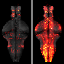
Whole-brain imaging allows for comprehensive functional mapping of distributed neural pathways, but neuronal perturbation experiments are usually limited to targeting predefined regions or genetically identifiable cell types. To complement whole-brain measures of activity with brain-wide manipulations for testing causal interactions, we introduce a system that uses measuredactivity patterns to guide optical perturbations of any subset of neurons in the same fictively behaving larval zebrafish. First, a light-sheet microscope collects whole-brain data that are rapidly analyzed by a distributed computing system to generate functional brain maps. On the basis of these maps, the experimenter can then optically ablate neurons and image activity changes across the brain. We applied this method to characterize contributions of behaviorally tuned populations to the optomotor response. We extended the system to optogenetically stimulate arbitrary subsets of neurons during whole-brain imaging. These open-source methods enable delineating the contributions of neurons to brain-wide circuit dynamics and behavior in individual animals.
In the absence of salient sensory cues to guide behavior, animals must still execute sequences of motor actions in order to forage and explore. How such successive motor actions are coordinated to form global locomotion trajectories is unknown. We mapped the structure of larval zebrafish swim trajectories in homogeneous environments and found that trajectories were characterized by alternating sequences of repeated turns to the left and to the right. Using whole-brain light-sheet imaging, we identified activity relating to the behavior in specific neural populations that we termed the anterior rhombencephalic turning region (ARTR). ARTR perturbations biased swim direction and reduced the dependence of turn direction on turn history, indicating that the ARTR is part of a network generating the temporal correlations in turn direction. We also find suggestive evidence for ARTR mutual inhibition and ARTR projections to premotor neurons. Finally, simulations suggest the observed turn sequences may underlie efficient exploration of local environments.
To investigate the fundamental question of how nervous systems encode, organize, and sequence behaviors, Kato et al. imaged neural activity with cellular resolution across the brain of the worm Caenorhabditis elegans. Locomotion behavior seems to be continuously represented by cyclical patterns of distributed neural activity that are present even in immobilized animals.
Understanding how the brain operates requires understanding how large sets of neurons function together. Modern recording technology makes it possible to simultaneously record the activity of hundreds of neurons, and technological developments will soon allow recording of thousands or tens of thousands. As with all experimental techniques, these methods are subject to confounds that complicate the interpretation of such recordings, and could lead to erroneous scientific conclusions. Here we discuss methods for assessing and improving the quality of data from these techniques and outline likely future directions in this field.
The processing of sensory input and the generation of behavior involves large networks of neurons, which necessitates new technology for recording from many neurons in behaving animals. In the larval zebrafish, light-sheet microscopy can be used to record the activity of almost all neurons in the brain simultaneously at single-cell resolution. Existing implementations, however, cannot be combined with visually driven behavior because the light sheet scans over the eye, interfering with presentation of controlled visual stimuli. Here we describe a system that overcomes the confounding eye stimulation through the use of two light sheets and combines whole-brain light-sheet imaging with virtual reality for fictively behaving larval zebrafish.
Developments in electrical and optical recording technology are scaling up the size of neuronal populations that can be monitored simultaneously. Light-sheet imaging is rapidly gaining traction as a method for optically interrogating activity in large networks and presents both opportunities and challenges for understanding circuit function.
Understanding brain function requires monitoring and interpreting the activity of large networks of neurons during behavior. Advances in recording technology are greatly increasing the size and complexity of neural data. Analyzing such data will pose a fundamental bottleneck for neuroscience. We present a library of analytical tools called Thunder built on the open-source Apache Spark platform for large-scale distributed computing. The library implements a variety of univariate and multivariate analyses with a modular, extendable structure well-suited to interactive exploration and analysis development. We demonstrate how these analyses find structure in large-scale neural data, including whole-brain light-sheet imaging data from fictively behaving larval zebrafish, and two-photon imaging data from behaving mouse. The analyses relate neuronal responses to sensory input and behavior, run in minutes or less and can be used on a private cluster or in the cloud. Our open-source framework thus holds promise for turning brain activity mapping efforts into biological insights.
The function of a neural circuit is shaped by the computations performed by its interneurons, which in many cases are not easily accessible to experimental investigation. Here, we elucidate the transformation of visual signals flowing from the input to the output of the primate retina, using a combination of large-scale multi-electrode recordings from an identified ganglion cell type, visual stimulation targeted at individual cone photoreceptors, and a hierarchical computational model. The results reveal nonlinear subunits in the circuity of OFF midget ganglion cells, which subserve high-resolution vision. The model explains light responses to a variety of stimuli more accurately than a linear model, including stimuli targeted to cones within and across subunits. The recovered model components are consistent with known anatomical organization of midget bipolar interneurons. These results reveal the spatial structure of linear and nonlinear encoding, at the resolution of single cells and at the scale of complete circuits.
Apache Spark is a popular open-source platform for large-scale data processing that is well-suited for iterative machine learning tasks. In this paper we present MLlib, Spark’s open-source distributed machine learning library. MLlib provides efficient functionality for a wide range of learning settings and includes several underlying statistical, optimization, and linear algebra primitives. Shipped with Spark, MLlib supports several languages and provides a high-level API that leverages Spark’s rich ecosystem to simplify the development of end-to-end machine learning pipelines. MLlib has experienced a rapid growth due to its vibrant open-source community of over 140 contributors, and includes extensive documentation to support further growth and to let users quickly get up to speed.