Filter
Associated Lab
- Branson Lab (3) Apply Branson Lab filter
- Card Lab (5) Apply Card Lab filter
- Dickson Lab (1) Apply Dickson Lab filter
- Jayaraman Lab (2) Apply Jayaraman Lab filter
- Remove Reiser Lab filter Reiser Lab
- Romani Lab (4) Apply Romani Lab filter
- Rubin Lab (15) Apply Rubin Lab filter
- Stern Lab (1) Apply Stern Lab filter
- Turaga Lab (2) Apply Turaga Lab filter
- Zuker Lab (1) Apply Zuker Lab filter
Associated Project Team
Publication Date
- 2024 (3) Apply 2024 filter
- 2023 (6) Apply 2023 filter
- 2022 (3) Apply 2022 filter
- 2021 (3) Apply 2021 filter
- 2020 (2) Apply 2020 filter
- 2019 (2) Apply 2019 filter
- 2018 (4) Apply 2018 filter
- 2017 (6) Apply 2017 filter
- 2016 (3) Apply 2016 filter
- 2015 (2) Apply 2015 filter
- 2014 (2) Apply 2014 filter
- 2013 (2) Apply 2013 filter
- 2012 (1) Apply 2012 filter
- 2011 (2) Apply 2011 filter
- 2010 (3) Apply 2010 filter
- 2009 (1) Apply 2009 filter
- 2008 (1) Apply 2008 filter
- 2007 (2) Apply 2007 filter
- 2003 (1) Apply 2003 filter
Type of Publication
49 Publications
Showing 41-49 of 49 resultsIt has long been known that many flying insects use visual cues to orient with respect to the wind and to control their groundspeed in the face of varying wind conditions. Much less explored has been the role of mechanosensory cues in orienting insects relative to the ambient air. Here we show that Drosophila melanogaster, magnetically tethered so as to be able to rotate about their yaw axis, are able to detect and orient into a wind, as would be experienced during forward flight. Further, this behavior is velocity dependent and is likely subserved, at least in part, by the Johnston’s organs, chordotonal organs in the antennae also involved in near-field sound detection. These wind-mediated responses may help to explain how flies are able to fly forward despite visual responses that might otherwise inhibit this behavior. Expanding visual stimuli, such as are encountered during forward flight, are the most potent aversive visual cues known for D. melanogaster flying in a tethered paradigm. Accordingly, tethered flies strongly orient towards a focus of contraction, a problematic situation for any animal attempting to fly forward. We show in this study that wind stimuli, transduced via mechanosensory means, can compensate for the aversion to visual expansion and thus may help to explain how these animals are indeed able to maintain forward flight.
Drosophila melanogaster is a model organism rich in genetic tools to manipulate and identify neural circuits involved in specific behaviors. Here we present a technique for two-photon calcium imaging in the central brain of head-fixed Drosophila walking on an air-supported ball. The ball’s motion is tracked at high resolution and can be treated as a proxy for the fly’s own movements. We used the genetically encoded calcium sensor, GCaMP3.0, to record from important elements of the motion-processing pathway, the horizontal-system lobula plate tangential cells (LPTCs) in the fly optic lobe. We presented motion stimuli to the tethered fly and found that calcium transients in horizontal-system neurons correlated with robust optomotor behavior during walking. Our technique allows both behavior and physiology in identified neurons to be monitored in a genetic model organism with an extensive repertoire of walking behaviors.
Nervous systems combine lower-level sensory signals to detect higher-order stimulus features critical to survival, such as the visual looming motion created by an imminent collision or approaching predator. Looming-sensitive neurons have been identified in diverse animal species. Different large-scale visual features such as looming often share local cues, which means loom-detecting neurons face the challenge of rejecting confounding stimuli. Here we report the discovery of an ultra-selective looming detecting neuron, lobula plate/lobula columnar, type II (LPLC2) in Drosophila, and show how its selectivity is established by radial motion opponency. In the fly visual system, directionally selective small-field neurons called T4 and T5 form a spatial map in the lobula plate, where they each terminate in one of four retinotopic layers, such that each layer responds to motion in a different cardinal direction. Single-cell anatomical analysis reveals that each arm of the LPLC2 cross-shaped primary dendrites ramifies in one of these layers and extends along that layer's preferred motion direction. In vivo calcium imaging demonstrates that, as their shape predicts, individual LPLC2 neurons respond strongly to outward motion emanating from the centre of the neuron's receptive field. Each dendritic arm also receives local inhibitory inputs directionally selective for inward motion opposing the excitation. This radial motion opponency generates a balance of excitation and inhibition that makes LPLC2 non-responsive to related patterns of motion such as contraction, wide-field rotation or luminance change. As a population, LPLC2 neurons densely cover visual space and terminate onto the giant fibre descending neurons, which drive the jump muscle motor neuron to trigger an escape take off. Our findings provide a mechanistic description of the selective feature detection that flies use to discern and escape looming threats.
As an animal translates through the world, its eyes will experience a radiating pattern of optic flow in which there is a focus of expansion directly in front and a focus of contraction behind. For flying fruit flies, recent experiments indicate that flies actively steer away from patterns of expansion. Whereas such a reflex makes sense for avoiding obstacles, it presents a paradox of sorts because an insect could not navigate stably through a visual scene unless it tolerated flight towards a focus of expansion during episodes of forward translation. One possible solution to this paradox is that a fly’s behavior might change such that it steers away from strong expansion, but actively steers towards weak expansion. In this study, we use a tethered flight arena to investigate the influence of stimulus strength on the magnitude and direction of turning responses to visual expansion in flies. These experiments indicate that the expansion-avoidance behavior is speed dependent. At slower speeds of expansion, flies exhibit an attraction to the focus of expansion, whereas the behavior transforms to expansion avoidance at higher speeds. Open-loop experiments indicate that this inversion of the expansion-avoidance response depends on whether or not the head is fixed to the thorax. The inversion of the expansion-avoidance response with stimulus strength has a clear manifestation under closed-loop conditions. Flies will actively orient towards a focus of expansion at low temporal frequency but steer away from it at high temporal frequency. The change in the response with temporal frequency does not require motion stimuli directly in front or behind the fly. Animals in which the stimulus was presented within 120 deg sectors on each side consistently steered towards expansion at low temporal frequency and steered towards contraction at high temporal frequency. A simple model based on an array of Hassenstein-Reichardt type elementary movement detectors suggests that the inversion of the expansion-avoidance reflex can explain the spatial distribution of straight flight segments and collision-avoidance saccades when flies fly freely within an open circular arena.
The ability of insects to learn and navigate to specific locations in the environment has fascinated naturalists for decades. The impressive navigational abilities of ants, bees, wasps and other insects demonstrate that insects are capable of visual place learning, but little is known about the underlying neural circuits that mediate these behaviours. Drosophila melanogaster (common fruit fly) is a powerful model organism for dissecting the neural circuitry underlying complex behaviours, from sensory perception to learning and memory. Drosophila can identify and remember visual features such as size, colour and contour orientation. However, the extent to which they use vision to recall specific locations remains unclear. Here we describe a visual place learning platform and demonstrate that Drosophila are capable of forming and retaining visual place memories to guide selective navigation. By targeted genetic silencing of small subsets of cells in the Drosophila brain, we show that neurons in the ellipsoid body, but not in the mushroom bodies, are necessary for visual place learning. Together, these studies reveal distinct neuroanatomical substrates for spatial versus non-spatial learning, and establish Drosophila as a powerful model for the study of spatial memories.
Visual projection neurons (VPNs) provide an anatomical connection between early visual processing and higher brain regions. Here we characterize lobula columnar (LC) cells, a class of Drosophila VPNs that project to distinct central brain structures called optic glomeruli. We anatomically describe 22 different LC types and show that, for several types, optogenetic activation in freely moving flies evokes specific behaviors. The activation phenotypes of two LC types closely resemble natural avoidance behaviors triggered by a visual loom. In vivo two-photon calcium imaging reveals that these LC types respond to looming stimuli, while another type does not, but instead responds to the motion of a small object. Activation of LC neurons on only one side of the brain can result in attractive or aversive turning behaviors depending on the cell type. Our results indicate that LC neurons convey information on the presence and location of visual features relevant for specific behaviors.
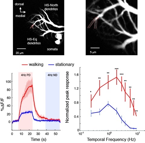
Changes in behavioral state modify neural activity in many systems. In some vertebrates such modulation has been observed and interpreted in the context of attention and sensorimotor coordinate transformations. Here we report state-dependent activity modulations during walking in a visual-motor pathway of Drosophila. We used two-photon imaging to monitor intracellular calcium activity in motion-sensitive lobula plate tangential cells (LPTCs) in head-fixed Drosophila walking on an air-supported ball. Cells of the horizontal system (HS)–a subgroup of LPTCs–showed stronger calcium transients in response to visual motion when flies were walking rather than resting. The amplified responses were also correlated with walking speed. Moreover, HS neurons showed a relatively higher gain in response strength at higher temporal frequencies, and their optimum temporal frequency was shifted toward higher motion speeds. Walking-dependent modulation of HS neurons in the Drosophila visual system may constitute a mechanism to facilitate processing of higher image speeds in behavioral contexts where these speeds of visual motion are relevant for course stabilization.
The body of an animal determines how the nervous system produces behavior. Therefore, detailed modeling of the neural control of sensorimotor behavior requires a detailed model of the body. Here we contribute an anatomically-detailed biomechanical whole-body model of the fruit fly Drosophila melanogaster in the MuJoCo physics engine. Our model is general-purpose, enabling the simulation of diverse fly behaviors, both on land and in the air. We demonstrate the generality of our model by simulating realistic locomotion, both flight and walking. To support these behaviors, we have extended MuJoCo with phenomenological models of fluid forces and adhesion forces. Through data-driven end-to-end reinforcement learning, we demonstrate that these advances enable the training of neural network controllers capable of realistic locomotion along complex trajectories based on high-level steering control signals. With a visually guided flight task, we demonstrate a neural controller that can use the vision sensors of the body model to control and steer flight. Our project is an open-source platform for modeling neural control of sensorimotor behavior in an embodied context.Competing Interest StatementThe authors have declared no competing interest.

An important strategy for efficient neural coding is to match the range of cellular responses to the distribution of relevant input signals. However, the structure and relevance of sensory signals depend on behavioral state. Here, we show that behavior modifies neural activity at the earliest stages of fly vision. We describe a class of wide-field neurons that provide feedback to the most peripheral layer of the Drosophila visual system, the lamina. Using in vivo patch-clamp electrophysiology, we found that lamina wide-field neurons respond to low-frequency luminance fluctuations. Recordings in flying flies revealed that the gain and frequency tuning of wide-field neurons change during flight, and that these effects are mimicked by the neuromodulator octopamine. Genetically silencing wide-field neurons increased behavioral responses to slow-motion stimuli. Together, these findings identify a cell type that is gated by behavior to enhance neural coding by subtracting low-frequency signals from the inputs to motion detection circuits.