Filter
Associated Lab
- Druckmann Lab (1) Apply Druckmann Lab filter
- Fitzgerald Lab (1) Apply Fitzgerald Lab filter
- Gonen Lab (1) Apply Gonen Lab filter
- Kainmueller Lab (1) Apply Kainmueller Lab filter
- Keller Lab (1) Apply Keller Lab filter
- Looger Lab (1) Apply Looger Lab filter
- Murphy Lab (1) Apply Murphy Lab filter
- Rubin Lab (1) Apply Rubin Lab filter
- Svoboda Lab (1) Apply Svoboda Lab filter
- Tjian Lab (1) Apply Tjian Lab filter
- Turner Lab (1) Apply Turner Lab filter
Publication Date
- August 24, 2011 (1) Apply August 24, 2011 filter
- August 23, 2011 (1) Apply August 23, 2011 filter
- August 19, 2011 (1) Apply August 19, 2011 filter
- August 17, 2011 (2) Apply August 17, 2011 filter
- August 16, 2011 (1) Apply August 16, 2011 filter
- August 9, 2011 (1) Apply August 9, 2011 filter
- August 2, 2011 (1) Apply August 2, 2011 filter
- August 1, 2011 (6) Apply August 1, 2011 filter
- Remove August 2011 filter August 2011
- Remove 2011 filter 2011
Type of Publication
14 Publications
Showing 1-10 of 14 resultsThe exact localization of the mandibular nerve with respect to the bone is important for applications in dental implantology and maxillofacial surgery. Cone beam computed tomography (CBCT), often also called digital volume tomography (DVT), is increasingly utilized in maxillofacial or dental imaging. Compared to conventional CT, however, soft tissue discrimination is worse due to a reduced dose. Thus, small structures like the alveolar nerves are even harder recognizable within the image data. We show that it is nonetheless possible to accurately reconstruct the 3D bone surface and the course of the nerve in a fully automatic fashion, with a method that is based on a combined statistical shape model of the nerve and the bone and a Dijkstra-based optimization procedure. Our method has been validated on 106 clinical datasets: the average reconstruction error for the bone is 0.5 +/- 0.1 mm, and the nerve can be detected with an average error of 1.0 +/- 0.6 mm.
Sensory stimuli are represented in the brain by the activity of populations of neurons. In most biological systems, studying population coding is challenging since only a tiny proportion of cells can be recorded simultaneously. Here we used two-photon imaging to record neural activity in the relatively simple Drosophila mushroom body (MB), an area involved in olfactory learning and memory. Using the highly sensitive calcium indicator GCaMP3, we simultaneously monitored the activity of >100 MB neurons in vivo (∼5% of the total population). The MB is thought to encode odors in sparse patterns of activity, but the code has yet to be explored either on a population level or with a wide variety of stimuli. We therefore imaged responses to odors chosen to evaluate the robustness of sparse representations. Different odors activated distinct patterns of MB neurons; however, we found no evidence for spatial organization of neurons by either response probability or odor tuning within the cell body layer. The degree of sparseness was consistent across a wide range of stimuli, from monomolecular odors to artificial blends and even complex natural smells. Sparseness was mainly invariant across concentrations, largely because of the influence of recent odor experience. Finally, in contrast to sensory processing in other systems, no response features distinguished natural stimuli from monomolecular odors. Our results indicate that the fundamental feature of odor processing in the MB is to create sparse stimulus representations in a format that facilitates arbitrary associations between odor and punishment or reward.
The rich dynamical nature of neurons poses major conceptual and technical challenges for unraveling their nonlinear membrane properties. Traditionally, various current waveforms have been injected at the soma to probe neuron dynamics, but the rationale for selecting specific stimuli has never been rigorously justified. The present experimental and theoretical study proposes a novel framework, inspired by learning theory, for objectively selecting the stimuli that best unravel the neuron’s dynamics. The efficacy of stimuli is assessed in terms of their ability to constrain the parameter space of biophysically detailed conductance-based models that faithfully replicate the neuron’s dynamics as attested by their ability to generalize well to the neuron’s response to novel experimental stimuli. We used this framework to evaluate a variety of stimuli in different types of cortical neurons, ages and animals. Despite their simplicity, a set of stimuli consisting of step and ramp current pulses outperforms synaptic-like noisy stimuli in revealing the dynamics of these neurons. The general framework that we propose paves a new way for defining, evaluating and standardizing effective electrical probing of neurons and will thus lay the foundation for a much deeper understanding of the electrical nature of these highly sophisticated and non-linear devices and of the neuronal networks that they compose.
Parallel circuits throughout the CNS exhibit distinct sensitivities and responses to sensory stimuli. Ambiguities in the source and properties of signals elicited by physiological stimuli, however, frequently obscure the mechanisms underlying these distinctions. We found that differences in the degree to which activity in two classes of Off retinal ganglion cell (RGC) encode information about light stimuli near detection threshold were not due to obvious differences in the cells’ intrinsic properties or the chemical synaptic input the cells received; indeed, differences in the cells’ light responses were largely insensitive to block of fast ionotropic glutamate receptors. Instead, the distinct responses of the two types of RGCs likely reflect differences in light-evoked electrical synaptic input. These results highlight a surprising strategy by which the retina differentially processes and routes visual information and provide new insight into the circuits that underlie responses to stimuli near detection threshold.
The ways in which cells set the size of intracellular structures is an important but largely unsolved problem [1]. Early embryonic divisions pose special problems in this regard. Many checkpoints common in somatic cells are missing from these divisions, which are characterized by rapid reductions in cell size and short cell cycles [2]. Embryonic cells must therefore possess simple and robust mechanisms that allow the size of many of their intracellular structures to rapidly scale with cell size.
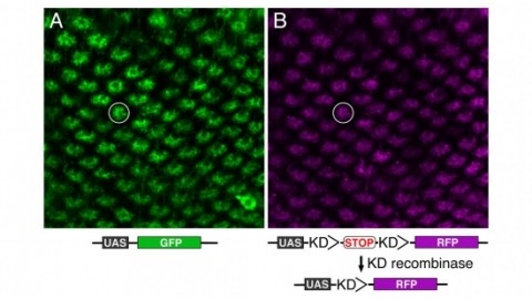
Site-specific recombinases have been used for two decades to manipulate the structure of animal genomes in highly predictable ways and have become major research tools. However, the small number of recombinases demonstrated to have distinct specificities, low toxicity, and sufficient activity to drive reactions to completion in animals has been a limitation. In this report we show that four recombinases derived from yeast-KD, B2, B3, and R-are highly active and nontoxic in Drosophila and that KD, B2, B3, and the widely used FLP recombinase have distinct target specificities. We also show that the KD and B3 recombinases are active in mice.
Bacterial Rho-independent terminators (RITs) are important genomic landmarks involved in gene regulation and terminating gene expression. In this investigation we present RNIE, a probabilistic approach for predicting RITs. The method is based upon covariance models which have been known for many years to be the most accurate computational tools for predicting homology in structural non-coding RNAs. We show that RNIE has superior performance in model species from a spectrum of bacterial phyla. Further analysis of species where a low number of RITs were predicted revealed a highly conserved structural sequence motif enriched near the genic termini of the pathogenic Actinobacteria, Mycobacterium tuberculosis. This motif, together with classical RITs, account for up to 90% of all the significantly structured regions from the termini of M. tuberculosis genic elements. The software, predictions and alignments described below are available from http://github.com/ppgardne/RNIE.
Secretins form megadalton bacterial-membrane channels in at least four sophisticated multiprotein systems that are crucial for translocation of proteins and assembled fibers across the outer membrane of many species of bacteria. Secretin subunits contain multiple domains, which interact with numerous other proteins, including pilotins, secretion-system partner proteins, and exoproteins. Our understanding of the structure of secretins is rapidly progressing, and it is now recognized that features common to all secretins include a cylindrical arrangement of 12-15 subunits, a large periplasmic vestibule with a wide opening at one end and a periplasmic gate at the other. Secretins might also play a key role in the biogenesis of their cognate secretion systems.
Pavlovian olfactory learning in Drosophila produces two genetically distinct forms of intermediate-term memories: anesthesia-sensitive memory, which requires the amnesiac gene, and anesthesia-resistant memory (ARM), which requires the radish gene. Here, we report that ARM is specifically enhanced or inhibited in flies with elevated or reduced serotonin (5HT) levels, respectively. The requirement for 5HT was additive with the memory defect of the amnesiac mutation but was occluded by the radish mutation. This result suggests that 5HT and Radish protein act on the same pathway for ARM formation. Three supporting lines of evidence indicate that ARM formation requires 5HT released from only two dorsal paired medial (DPM) neurons onto the mushroom bodies (MBs), the olfactory learning and memory center in Drosophila: (i) DPM neurons were 5HT-antibody immunopositive; (ii) temporal inhibition of 5HT synthesis or release from DPM neurons, but not from other serotonergic neurons, impaired ARM formation; (iii) knocking down the expression of d5HT1A serotonin receptors in α/β MB neurons, which are innervated by DPM neurons, inhibited ARM formation. Thus, in addition to the Amnesiac peptide required for anesthesia-sensitive memory formation, the two DPM neurons also release 5HT acting on MB neurons for ARM formation.

Light sheet-based fluorescence microscopy (LSFM) is emerging as a powerful imaging technique for the life sciences. LSFM provides an exceptionally high imaging speed, high signal-to-noise ratio, low level of photo-bleaching and good optical penetration depth. This unique combination of capabilities makes light sheet-based microscopes highly suitable for live imaging applications. There is an outstanding potential in applying this technology to the quantitative study of embryonic development. Here, we provide an overview of the different basic implementations of LSFM, review recent technical advances in the field and highlight applications in the context of embryonic development. We conclude with a discussion of promising future directions.