Filter
Associated Lab
- Ahrens Lab (3) Apply Ahrens Lab filter
- Betzig Lab (8) Apply Betzig Lab filter
- Card Lab (2) Apply Card Lab filter
- Cardona Lab (1) Apply Cardona Lab filter
- Druckmann Lab (1) Apply Druckmann Lab filter
- Eddy/Rivas Lab (2) Apply Eddy/Rivas Lab filter
- Fetter Lab (4) Apply Fetter Lab filter
- Fitzgerald Lab (1) Apply Fitzgerald Lab filter
- Gonen Lab (6) Apply Gonen Lab filter
- Harris Lab (1) Apply Harris Lab filter
- Heberlein Lab (3) Apply Heberlein Lab filter
- Hess Lab (2) Apply Hess Lab filter
- Ji Lab (4) Apply Ji Lab filter
- Kainmueller Lab (1) Apply Kainmueller Lab filter
- Keller Lab (3) Apply Keller Lab filter
- Lavis Lab (4) Apply Lavis Lab filter
- Looger Lab (3) Apply Looger Lab filter
- Magee Lab (4) Apply Magee Lab filter
- Murphy Lab (1) Apply Murphy Lab filter
- Pastalkova Lab (3) Apply Pastalkova Lab filter
- Reiser Lab (1) Apply Reiser Lab filter
- Riddiford Lab (5) Apply Riddiford Lab filter
- Romani Lab (2) Apply Romani Lab filter
- Rubin Lab (4) Apply Rubin Lab filter
- Saalfeld Lab (2) Apply Saalfeld Lab filter
- Satou Lab (1) Apply Satou Lab filter
- Schreiter Lab (3) Apply Schreiter Lab filter
- Sgro Lab (1) Apply Sgro Lab filter
- Shroff Lab (6) Apply Shroff Lab filter
- Spruston Lab (3) Apply Spruston Lab filter
- Stern Lab (4) Apply Stern Lab filter
- Sternson Lab (1) Apply Sternson Lab filter
- Svoboda Lab (6) Apply Svoboda Lab filter
- Tjian Lab (7) Apply Tjian Lab filter
- Truman Lab (2) Apply Truman Lab filter
- Turner Lab (1) Apply Turner Lab filter
- Wu Lab (1) Apply Wu Lab filter
- Zuker Lab (1) Apply Zuker Lab filter
Publication Date
- Remove 2007-12-31 19:00 – 2008-12-31 19:00 filter 2007-12-31 19:00 – 2008-12-31 19:00
- December 2008 (11) Apply December 2008 filter
- November 2008 (8) Apply November 2008 filter
- October 2008 (7) Apply October 2008 filter
- September 2008 (12) Apply September 2008 filter
- August 2008 (10) Apply August 2008 filter
- July 2008 (14) Apply July 2008 filter
- June 2008 (13) Apply June 2008 filter
- May 2008 (8) Apply May 2008 filter
- April 2008 (7) Apply April 2008 filter
- March 2008 (16) Apply March 2008 filter
- February 2008 (13) Apply February 2008 filter
- January 2008 (21) Apply January 2008 filter
Type of Publication
140 Publications
Showing 51-60 of 140 resultsMany important physiological processes operate at time and space scales far beyond those accessible to atom-realistic simulations, and yet discrete stochastic rather than continuum methods may best represent finite numbers of molecules interacting in complex cellular spaces. We describe and validate new tools and algorithms developed for a new version of the MCell simulation program (MCell3), which supports generalized Monte Carlo modeling of diffusion and chemical reaction in solution, on surfaces representing membranes, and combinations thereof. A new syntax for describing the spatial directionality of surface reactions is introduced, along with optimizations and algorithms that can substantially reduce computational costs (e.g., event scheduling, variable time and space steps). Examples for simple reactions in simple spaces are validated by comparison to analytic solutions. Thus we show how spatially realistic Monte Carlo simulations of biological systems can be far more cost-effective than often is assumed, and provide a level of accuracy and insight beyond that of continuum methods.
In order to study anatomy of organisms with high-resolution there is an increasing demand to image large specimen in three dimensions (3D). Confocal microscopy is able to produce high-resolution 3D images, but these are limited by its relatively small field of view compared to the size of large biological specimens. To overcome this drawback, motorized stages moving the sample are used to create a tiled scan of the whole specimen. The physical coordinates provided by the microscope stage are not precise enough to allow reconstruction (”Stitching”) of the whole image from individual image stacks.
We developed an algorithm, as well as an ImageJ plug-in, based on the Fourier Shift Theorem that computes all possible translations (x, y, z) between two 3D images at once, yielding the best overlap in terms of the cross correlation measure. Apart from the obvious gain in computation time it has the advantage that it cannot be trapped in local minima as it simply computes all possible solutions. Computing the overlap between two adjacent image stacks is fast (12 seconds for two 512x512x89 images on a Intel ® Core2Duo with 2.2GHz) making it suitable for real time use, i.e. computing the output image during acquisition of the individual image stacks.
To compensate the possible shading- and brightness differences we apply a smooth linear intensity transition between the overlapping stacks. Additionally we extended the to generic 3D registration using gradient based rotation detection on top of the phase correlation method. We demonstrate the performance of our 3D stitching plug-in on several tiled confocal images and show an example of its application for 3D registration.
Understanding the principles of information processing in neural circuits requires systematic characterization of the participating cell types and their connections, and the ability to measure and perturb their activity. Genetic approaches promise to bring experimental access to complex neural systems, including genetic stalwarts such as the fly and mouse, but also to nongenetic systems such as primates. Together with anatomical and physiological methods, cell-type-specific expression of protein markers and sensors and transducers will be critical to construct circuit diagrams and to measure the activity of genetically defined neurons. Inactivation and activation of genetically defined cell types will establish causal relationships between activity in specific groups of neurons, circuit function, and animal behavior. Genetic analysis thus promises to reveal the logic of the neural circuits in complex brains that guide behaviors. Here we review progress in the genetic analysis of neural circuits and discuss directions for future research and development.
The ad hoc genetic correlation between ethanol sensitivity and learning mechanisms in Drosophila could overemphasize a common process supporting both behaviors. To challenge directly the hypothesis that these mechanisms are singular, we examined the learning phenotypes of 10 new strains. Five of these have increased ethanol sensitivity, and the other 5 do not. We tested place and olfactory memory in each of these lines and found two new learning mutations. In one case, altering the tribbles gene, flies have a significantly reduced place memory, elevated olfactory memory, and normal ethanol response. In the second case, mutation of a gene we name ethanol sensitive with low memory (elm), place memory was not altered, olfactory memory was sharply reduced, and sensitivity to ethanol was increased. In sum, however, we found no overall correlation between ethanol sensitivity and place memory in the 10 lines tested. Furthermore, there was a weak but nonsignificant correlation between ethanol sensitivity and olfactory learning. Thus, mutations that alter learning and sensitivity to ethanol can occur independently of each other and this implies that the set of genes important for both ethanol sensitivity and learning is likely a subset of the genes important for either process.
Neurons and glia are functionally organized into circuits and higher-order structures via synaptic connectivity, well-orchestrated molecular signaling, and activity-dependent refinement. Such organization allows the precise information processing required for complex behaviors. Disruption of nervous systems by genetic deficiency or events such as trauma or environmental exposure may produce a diseased state in which certain aspects of inter-neuron signaling are impaired. Optical imaging techniques allow the direct visualization of individual neurons in a circuit environment. Imaging probes specific for given biomolecules may help elucidate their contribution to proper circuit function. Genetically encoded sensors can visualize trafficking of particular molecules in defined neuronal populations, non-invasively in intact brain or reduced preparations. Sensor analysis in healthy and diseased brains may reveal important differences and shed light on the development and progression of nervous system disorders. We review the field of genetically encoded sensors for molecules and cellular events, and their potential applicability to the study of nervous system disease.
Drosophila mushroom body (MB) gamma neurons undergo axon pruning during metamorphosis through a process of localized degeneration of specific axon branches. Developmental axon degeneration is initiated by the steroid hormone ecdysone, acting through a nuclear receptor complex composed of USP (ultraspiracle) and EcRB1 (ecdysone receptor B1) to regulate gene expression in MB gamma neurons. To identify ecdysone-dependent gene expression changes in MB gamma neurons at the onset of axon pruning, we use laser capture microdissection to isolate wild-type and mutant MB neurons in which EcR (ecdysone receptor) activity is genetically blocked, and analyze expression changes by microarray. We identify several molecular pathways that are regulated in MB neurons by ecdysone. The most striking observation is the upregulation of genes involved in the UPS (ubiquitin-proteasome system), which is cell autonomously required for gamma neuron pruning. In addition, we characterize the function of Boule, an evolutionarily conserved RNA-binding protein previously implicated in spermatogenesis in flies and vertebrates. boule expression is downregulated by ecdysone in MB neurons at the onset of pruning, and forced expression of Boule in MB gamma neurons is sufficient to inhibit axon pruning. This activity is dependent on the RNA-binding domain of Boule and a conserved DAZ (deleted in azoospermia) domain implicated in interactions with other RNA-binding proteins. However, loss of Boule does not result in obvious defects in axon pruning or morphogenesis of MB neurons, suggesting that it acts redundantly with other ecdyonse-regulated genes. We propose a novel function for Boule in the CNS as a negative regulator of developmental axon pruning.
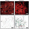
The identification of synaptic partners is challenging in dense nerve bundles, where many processes occupy regions beneath the resolution of conventional light microscopy. To address this difficulty, we have developed GRASP, a system to label membrane contacts and synapses between two cells in living animals. Two complementary fragments of GFP are expressed on different cells, tethered to extracellular domains of transmembrane carrier proteins. When the complementary GFP fragments are fused to ubiquitous transmembrane proteins, GFP fluorescence appears uniformly along membrane contacts between the two cells. When one or both GFP fragments are fused to synaptic transmembrane proteins, GFP fluorescence is tightly localized to synapses. GRASP marks known synaptic contacts in C. elegans, correctly identifies changes in mutants with altered synaptic specificity, and can uncover new information about synaptic locations as confirmed by electron microscopy. GRASP may prove particularly useful for defining connectivity in complex nervous systems.
We present a compressed domain scheme that is able to recognize and localize actions at high speeds. The recognition problem is posed as performing an action video query on a test video sequence. Our method is based on computing motion similarity using compressed domain features which can be extracted with low complexity. We introduce a novel motion correlation measure that takes into account differences in motion directions and magnitudes. Our method is appearance invariant, requires no prior segmentation, alignment or stabilization, and is able to localize actions in both space and time. We evaluated our method on a benchmark action video database consisting of 6 actions performed by 25 people under 3 different scenarios. Our proposed method achieved a classification accuracy of 90%, comparing favorably with existing methods in action classification accuracy, and is able to localize a template video of 80 x 64 pixels with 23 frames in a test video of 368 x 184 pixels with 835 frames in just 11 seconds, easily outperforming other methods in localization speed. We also perform a systematic investigation of the effects of various encoding options on our proposed approach. In particular, we present results on the compression-classification trade-off, which would provide valuable insight into jointly designing a system that performs video encoding at the camera front-end and action classification at the processing backend.
We combined photoactivated localization microscopy (PALM) with live-cell single-particle tracking to create a new method termed sptPALM. We created spatially resolved maps of single-molecule motions by imaging the membrane proteins Gag and VSVG, and obtained several orders of magnitude more trajectories per cell than traditional single-particle tracking enables. By probing distinct subsets of molecules, sptPALM can provide insight into the origins of spatial and temporal heterogeneities in membranes.
Commentary: As a stepping stone to true live cell PALM (see above), our collaborator Jennifer Lippincott-Schwartz suggested using the sparse photoactivation principle of PALM to track the nanoscale motion of thousands of individual molecules within a single living cell. Termed single particle tracking PALM (sptPALM), Jennifer’s postdocs Suliana Manley and Jen Gillette used the method in our PALM rig to create spatially resolved maps of diffusion rates in the plasma membrane of live cells. sptPALM is a powerful tool to study the active cytoskeletal or passive diffusional transport of individual molecules with far more measurements per cell than is possible without sparse photoactivation.
Pulsed lasers are key elements in nonlinear bioimaging techniques such as two-photon fluorescence excitation (TPE) microscopy. Typically, however, only a percent or less of the laser power available can be delivered to the sample before photoinduced damage becomes excessive. Here we describe a passive pulse splitter that converts each laser pulse into a fixed number of sub-pulses of equal energy. We applied the splitter to TPE imaging of fixed mouse brain slices labeled with GFP and show that, in different power regimes, the splitter can be used either to increase the signal rate more than 100-fold or to reduce the rate of photobleaching by over fourfold. In living specimens, the gains were even greater: a ninefold reduction in photobleaching during in vivo imaging of Caenorhabditis elegans larvae, and a six- to 20-fold decrease in the rate of photodamage during calcium imaging of rat hippocampal brain slices.