Filter
Associated Lab
- Ahrens Lab (6) Apply Ahrens Lab filter
- Betzig Lab (1) Apply Betzig Lab filter
- Dudman Lab (1) Apply Dudman Lab filter
- Fitzgerald Lab (1) Apply Fitzgerald Lab filter
- Harris Lab (5) Apply Harris Lab filter
- Heberlein Lab (1) Apply Heberlein Lab filter
- Hermundstad Lab (1) Apply Hermundstad Lab filter
- Jayaraman Lab (7) Apply Jayaraman Lab filter
- Ji Lab (3) Apply Ji Lab filter
- Karpova Lab (1) Apply Karpova Lab filter
- Lavis Lab (1) Apply Lavis Lab filter
- Leonardo Lab (2) Apply Leonardo Lab filter
- Looger Lab (16) Apply Looger Lab filter
- Podgorski Lab (4) Apply Podgorski Lab filter
- Rubin Lab (1) Apply Rubin Lab filter
- Schreiter Lab (19) Apply Schreiter Lab filter
- Svoboda Lab (13) Apply Svoboda Lab filter
- Tillberg Lab (1) Apply Tillberg Lab filter
- Turner Lab (2) Apply Turner Lab filter
- Zlatic Lab (1) Apply Zlatic Lab filter
Associated Project Team
Publication Date
- 2024 (3) Apply 2024 filter
- 2023 (5) Apply 2023 filter
- 2022 (1) Apply 2022 filter
- 2020 (5) Apply 2020 filter
- 2019 (2) Apply 2019 filter
- 2018 (5) Apply 2018 filter
- 2017 (3) Apply 2017 filter
- 2016 (2) Apply 2016 filter
- 2015 (4) Apply 2015 filter
- 2014 (3) Apply 2014 filter
- 2013 (5) Apply 2013 filter
- 2012 (2) Apply 2012 filter
Type of Publication
- Remove Janelia filter Janelia
40 Publications
Showing 21-30 of 40 resultsFrom patch-clamp techniques to recombinant DNA technologies, three-dimensional protein modeling, and optogenetics, diverse and sophisticated methods have been used to study ion channels and how they determine the electrical properties of cells.
Femtosecond lasers at fixed wavelengths above 1,000 nm are powerful, stable and inexpensive, making them promising sources for two-photon microscopy. Biosensors optimized for these wavelengths are needed for both next-generation microscopes and affordable turn-key systems. Here we report jYCaMP1, a yellow variant of the calcium indicator jGCaMP7 that outperforms its parent in mice and flies at excitation wavelengths above 1,000 nm and enables improved two-color calcium imaging with red fluorescent protein-based indicators.
The identification of active neurons and circuits in vivo is a fundamental challenge in understanding the neural basis of behavior. Genetically encoded calcium (Ca(2+)) indicators (GECIs) enable quantitative monitoring of cellular-resolution activity during behavior. However, such indicators require online monitoring within a limited field of view. Alternatively, post hoc staining of immediate early genes (IEGs) indicates highly active cells within the entire brain, albeit with poor temporal resolution. We designed a fluorescent sensor, CaMPARI, that combines the genetic targetability and quantitative link to neural activity of GECIs with the permanent, large-scale labeling of IEGs, allowing a temporally precise "activity snapshot" of a large tissue volume. CaMPARI undergoes efficient and irreversible green-to-red conversion only when elevated intracellular Ca(2+) and experimenter-controlled illumination coincide. We demonstrate the utility of CaMPARI in freely moving larvae of zebrafish and flies, and in head-fixed mice and adult flies.
Recent success in training artificial agents and robots derives from a combination of direct learning of behavioural policies and indirect learning through value functions. Policy learning and value learning use distinct algorithms that optimize behavioural performance and reward prediction, respectively. In animals, behavioural learning and the role of mesolimbic dopamine signalling have been extensively evaluated with respect to reward prediction; however, so far there has been little consideration of how direct policy learning might inform our understanding. Here we used a comprehensive dataset of orofacial and body movements to understand how behavioural policies evolved as naive, head-restrained mice learned a trace conditioning paradigm. Individual differences in initial dopaminergic reward responses correlated with the emergence of learned behavioural policy, but not the emergence of putative value encoding for a predictive cue. Likewise, physiologically calibrated manipulations of mesolimbic dopamine produced several effects inconsistent with value learning but predicted by a neural-network-based model that used dopamine signals to set an adaptive rate, not an error signal, for behavioural policy learning. This work provides strong evidence that phasic dopamine activity can regulate direct learning of behavioural policies, expanding the explanatory power of reinforcement learning models for animal learning.
We describe an adaptive optics method that modulates the intensity or phase of light rays at multiple pupil segments in parallel to determine the sample-induced aberration. Applicable to fluorescent protein-labeled structures of arbitrary complexity, it allowed us to obtain diffraction-limited resolution in various samples in vivo. For the strongly scattering mouse brain, a single aberration correction improved structural and functional imaging of fine neuronal processes over a large imaging volume.
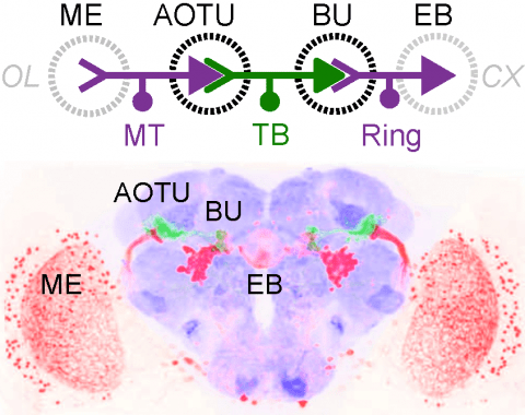
Many animals orient using visual cues, but how a single cue is selected from among many is poorly understood. Here we show that Drosophila ring neurons—central brain neurons implicated in navigation—display visual stimulus selection. Using in vivo two-color two-photon imaging with genetically encoded calcium indicators, we demonstrate that individual ring neurons inherit simple-cell-like receptive fields from their upstream partners. Stimuli in the contralateral visual field suppressed responses to ipsilateral stimuli in both populations. Suppression strength depended on when and where the contralateral stimulus was presented, an effect stronger in ring neurons than in their upstream inputs. This history-dependent effect on the temporal structure of visual responses, which was well modeled by a simple biphasic filter, may determine how visual references are selected for the fly's internal compass. Our approach highlights how two-color calcium imaging can help identify and localize the origins of sensory transformations across synaptically connected neural populations.
The mouse has become an important model for understanding the neural basis of visual perception. Although it has long been known that mouse lens transmits ultraviolet (UV) light and mouse opsins have absorption in the UV band, little is known about how UV visual information is processed in the mouse brain. Using a custom UV stimulation system and in vivo calcium imaging, we characterized the feature selectivity of layer 2/3 neurons in mouse primary visual cortex (V1). In adult mice, a comparable percentage of the neuronal population responds to UV and visible stimuli, with similar pattern selectivity and receptive field properties. In young mice, the orientation selectivity for UV stimuli increased steadily during development, but not direction selectivity. Our results suggest that, by expanding the spectral window through which the mouse can acquire visual information, UV sensitivity provides an important component for mouse vision.
Temporal focusing (TF) multiphoton systems constitute a powerful solution for cellular resolution optogenetic stimulation and recording in three-dimensional, scattering tissue. Here, we address two fundamental aspects in the design of such systems: first, we examine the design of TF systems with specific optical sectioning by comparatively analyzing previously published results. Next, we develop a solution for obtaining TF in a flexible three-dimensional pattern of cellmatched focal spots. Our solution employs spatio-temporal focusing (SSTF) in a unique optical system design that can be integrated before essentially any multiphoton imaging or stimulation system.
Genetically encoded calcium indicators (GECIs) are powerful tools for systems neuroscience. Recent efforts in protein engineering have significantly increased the performance of GECIs. The state-of-the art single-wavelength GECI, GCaMP3, has been deployed in a number of model organisms and can reliably detect three or more action potentials in short bursts in several systems in vivo . Through protein structure determination, targeted mutagenesis, high-throughput screening, and a battery of in vitro assays, we have increased the dynamic range of GCaMP3 by severalfold, creating a family of “GCaMP5” sensors. We tested GCaMP5s in several systems: cultured neurons and astrocytes, mouse retina, and in vivo in Caenorhabditis chemosensory neurons, Drosophila larval neuromuscular junction and adult antennal lobe, zebrafish retina and tectum, and mouse visual cortex. Signal-to-noise ratio was improved by at least 2- to 3-fold. In the visual cortex, two GCaMP5 variants detected twice as many visual stimulus-responsive cells as GCaMP3. By combining in vivo imaging with electrophysiology we show that GCaMP5 fluorescence provides a more reliable measure of neuronal activity than its predecessor GCaMP3.GCaMP5allows more sensitive detection of neural activity in vivo andmayfind widespread applications for cellular imaging in general.
The quality of genetically encoded calcium indicators (GECIs) has improved dramatically in recent years, but high-performing ratiometric indicators are still rare. Here we describe a series of fluorescence resonance energy transfer (FRET)-based calcium biosensors with a reduced number of calcium binding sites per sensor. These ’Twitch’ sensors are based on the C-terminal domain of Opsanus troponin C. Their FRET responses were optimized by a large-scale functional screen in bacterial colonies, refined by a secondary screen in rat hippocampal neuron cultures. We tested the in vivo performance of the most sensitive variants in the brain and lymph nodes of mice. The sensitivity of the Twitch sensors matched that of synthetic calcium dyes and allowed visualization of tonic action potential firing in neurons and high resolution functional tracking of T lymphocytes. Given their ratiometric readout, their brightness, large dynamic range and linear response properties, Twitch sensors represent versatile tools for neuroscience and immunology.

