Filter
Associated Lab
- Aso Lab (1) Apply Aso Lab filter
- Branson Lab (1) Apply Branson Lab filter
- Card Lab (4) Apply Card Lab filter
- Cardona Lab (17) Apply Cardona Lab filter
- Dickson Lab (1) Apply Dickson Lab filter
- Fetter Lab (9) Apply Fetter Lab filter
- Heberlein Lab (1) Apply Heberlein Lab filter
- Riddiford Lab (7) Apply Riddiford Lab filter
- Rubin Lab (4) Apply Rubin Lab filter
- Simpson Lab (2) Apply Simpson Lab filter
- Singer Lab (1) Apply Singer Lab filter
- Stern Lab (4) Apply Stern Lab filter
- Remove Truman Lab filter Truman Lab
- Zlatic Lab (13) Apply Zlatic Lab filter
Associated Project Team
Associated Support Team
Publication Date
- 2023 (2) Apply 2023 filter
- 2021 (3) Apply 2021 filter
- 2020 (3) Apply 2020 filter
- 2019 (4) Apply 2019 filter
- 2018 (8) Apply 2018 filter
- 2017 (6) Apply 2017 filter
- 2016 (11) Apply 2016 filter
- 2015 (6) Apply 2015 filter
- 2014 (1) Apply 2014 filter
- 2013 (3) Apply 2013 filter
- 2012 (3) Apply 2012 filter
- 2011 (1) Apply 2011 filter
- 2010 (4) Apply 2010 filter
- 2009 (2) Apply 2009 filter
- 2008 (1) Apply 2008 filter
58 Janelia Publications
Showing 51-58 of 58 resultsThe coordination of growth with nutritional status is essential for proper development and physiology. Nutritional information is mostly perceived by peripheral organs before being relayed to the brain, which modulates physiological responses. Hormonal signaling ensures this organ-to-organ communication, and the failure of endocrine regulation in humans can cause diseases including obesity and diabetes. In Drosophila melanogaster, the fat body (adipose tissue) has been suggested to play an important role in coupling growth with nutritional status. Here, we show that the peripheral tissue-derived peptide hormone CCHamide-2 (CCHa2) acts as a nutrient-dependent regulator of Drosophila insulin-like peptides (Dilps). A BAC-based transgenic reporter revealed strong expression of CCHa2 receptor (CCHa2-R) in insulin-producing cells (IPCs) in the brain. Calcium imaging of brain explants and IPC-specific CCHa2-R knockdown demonstrated that peripheral-tissue derived CCHa2 directly activates IPCs. Interestingly, genetic disruption of either CCHa2 or CCHa2-R caused almost identical defects in larval growth and developmental timing. Consistent with these phenotypes, the expression of dilp5, and the release of both Dilp2 and Dilp5, were severely reduced. Furthermore, transcription of CCHa2 is altered in response to nutritional levels, particularly of glucose. These findings demonstrate that CCHa2 and CCHa2-R form a direct link between peripheral tissues and the brain, and that this pathway is essential for the coordination of systemic growth with nutritional availability. A mammalian homologue of CCHa2-R, Bombesin receptor subtype-3 (Brs3), is an orphan receptor that is expressed in the islet β-cells; however, the role of Brs3 in insulin regulation remains elusive. Our genetic approach in Drosophila melanogaster provides the first evidence, to our knowledge, that bombesin receptor signaling with its endogenous ligand promotes insulin production.
Mapping brain function to brain structure is a fundamental task for neuroscience. For such an endeavour, the Drosophila larva is simple enough to be tractable, yet complex enough to be interesting. It features about 10,000 neurons and is capable of various taxes, kineses and Pavlovian conditioning. All its neurons are currently being mapped into a light-microscopical atlas, and Gal4 strains are being generated to experimentally access neurons one at a time. In addition, an electron microscopic reconstruction of its nervous system seems within reach. Notably, this electron microscope-based connectome is being drafted for a stage 1 larva - because stage 1 larvae are much smaller than stage 3 larvae. However, most behaviour analyses have been performed for stage 3 larvae because their larger size makes them easier to handle and observe. It is therefore warranted to either redo the electron microscopic reconstruction for a stage 3 larva or to survey the behavioural faculties of stage 1 larvae. We provide the latter. In a community-based approach we called the Ol1mpiad, we probed stage 1 Drosophila larvae for free locomotion, feeding, responsiveness to substrate vibration, gentle and nociceptive touch, burrowing, olfactory preference and thermotaxis, light avoidance, gustatory choice of various tastants plus odour-taste associative learning, as well as light/dark-electric shock associative learning. Quantitatively, stage 1 larvae show lower scores in most tasks, arguably because of their smaller size and lower speed. Qualitatively, however, stage 1 larvae perform strikingly similar to stage 3 larvae in almost all cases. These results bolster confidence in mapping brain structure and behaviour across developmental stages.
The sense of smell enables animals to react to long-distance cues according to learned and innate valences. Here, we have mapped with electron microscopy the complete wiring diagram of the Drosophila larval antennal lobe, an olfactory neuropil similar to the vertebrate olfactory bulb. We found a canonical circuit with uniglomerular projection neurons (uPNs) relaying gain-controlled ORN activity to the mushroom body and the lateral horn. A second, parallel circuit with multiglomerular projection neurons (mPNs) and hierarchically connected local neurons (LNs) selectively integrates multiple ORN signals already at the first synapse. LN-LN synaptic connections putatively implement a bistable gain control mechanism that either computes odor saliency through panglomerular inhibition, or allows some glomeruli to respond to faint aversive odors in the presence of strong appetitive odors. This complete wiring diagram will support experimental and theoretical studies towards bridging the gap between circuits and behavior.
Higher-order genome organization plays an important role in transcriptional regulation. In Drosophila, somatic pairing of homologous chromosomes can lead to transvection, by which the regulatory region of a gene can influence transcription in trans. We observe transvection between transgenes inserted at commonly used phiC31 integration sites in the Drosophila genome. When two transgenes that carry endogenous regulatory elements driving the expression of either LexA or GAL4 are inserted at the same integration site and paired, the enhancer of one transgene can drive or repress expression of the paired transgene. These transvection effects depend on compatibility between regulatory elements and are often restricted to a subset of cell types within a given expression pattern. We further show that activated UAS-transgenes can also drive transcription in trans. We discuss the implication of these findings for 1) understanding the molecular mechanisms that underlie transvection and 2) the design of experiments that utilize site-specific integration.
BACKGROUND: In holometabolous insects such as Drosophila melanogaster, neuroblasts produce an initial population of diverse neurons during embryogenesis and a much larger set of adult-specific neurons during larval life. In the ventral CNS, many of these secondary neuronal lineages differ significantly from one body segment to another, suggesting a role for anteroposterior patterning genes. RESULTS: Here we systematically characterize the expression pattern and function of the Hox gene Ultrabithorax (Ubx) in all 25 postembryonic lineages. We find that Ubx is expressed in a segment-, lineage-, and hemilineage-specific manner in the thoracic and anterior abdominal segments. When Ubx is removed from neuroblasts via mitotic recombination, neurons in these segments exhibit the morphologies and survival patterns of their anterior thoracic counterparts. Conversely, when Ubx is ectopically expressed in anterior thoracic segments, neurons exhibit complementary posterior transformation phenotypes. CONCLUSION: Our findings demonstrate that Ubx plays a critical role in conferring segment-appropriate morphology and survival on individual neurons in the adult-specific ventral CNS. Moreover, while always conferring spatial identity in some sense, Ubx has been co-opted during evolution for distinct and even opposite functions in different neuronal hemilineages.
Neuroendocrine systems in animals maintain organismal homeostasis and regulate stress response. Although a great deal of work has been done on the neuropeptides and hormones that are released and act on target organs in the periphery, the synaptic inputs onto these neuroendocrine outputs in the brain are less well understood. Here, we use the transmission electron microscopy reconstruction of a whole central nervous system in the larva to elucidate the sensory pathways and the interneurons that provide synaptic input to the neurosecretory cells projecting to the endocrine organs. Predicted by network modeling, we also identify a new carbon dioxide-responsive network that acts on a specific set of neurosecretory cells and that includes those expressing corazonin (Crz) and diuretic hormone 44 (Dh44) neuropeptides. Our analysis reveals a neuronal network architecture for combinatorial action based on sensory and interneuronal pathways that converge onto distinct combinations of neuroendocrine outputs.
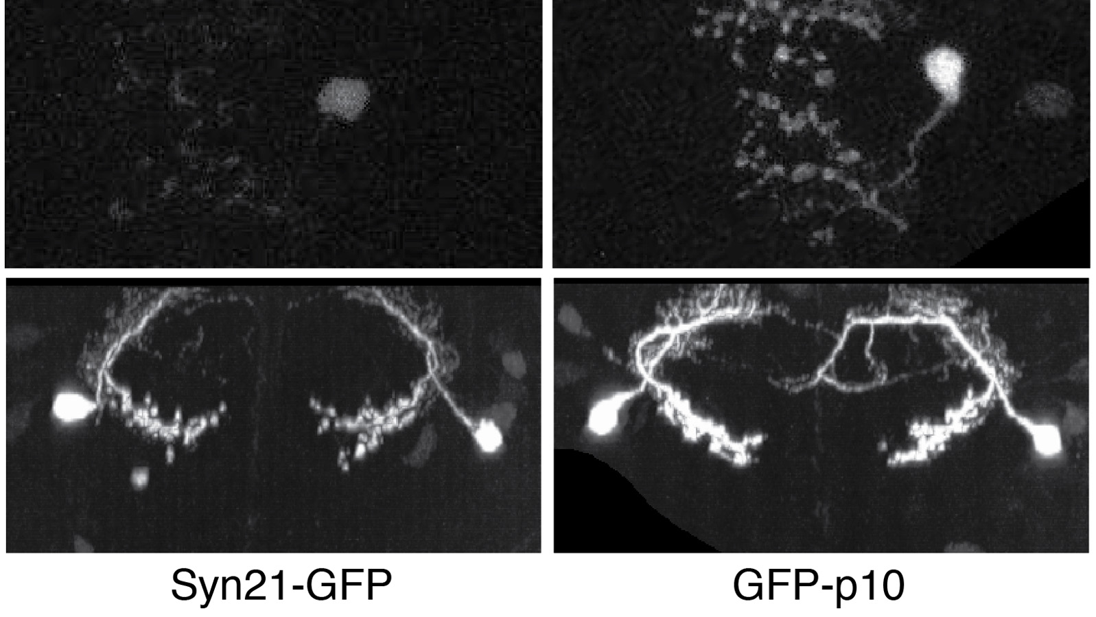
The ability to specify the expression levels of exogenous genes inserted in the genomes of transgenic animals is critical for the success of a wide variety of experimental manipulations. Protein production can be regulated at the level of transcription, mRNA transport, mRNA half-life, or translation efficiency. In this report, we show that several well-characterized sequence elements derived from plant and insect viruses are able to function in Drosophila to increase the apparent translational efficiency of mRNAs by as much as 20-fold. These increases render expression levels sufficient for genetic constructs previously requiring multiple copies to be effective in single copy, including constructs expressing the temperature-sensitive inactivator of neuronal function Shibire(ts1), and for the use of cytoplasmic GFP to image the fine processes of neurons.