Filter
Associated Lab
- Aguilera Castrejon Lab (1) Apply Aguilera Castrejon Lab filter
- Ahrens Lab (45) Apply Ahrens Lab filter
- Aso Lab (39) Apply Aso Lab filter
- Baker Lab (19) Apply Baker Lab filter
- Betzig Lab (98) Apply Betzig Lab filter
- Beyene Lab (4) Apply Beyene Lab filter
- Bock Lab (14) Apply Bock Lab filter
- Branson Lab (45) Apply Branson Lab filter
- Card Lab (34) Apply Card Lab filter
- Cardona Lab (44) Apply Cardona Lab filter
- Chklovskii Lab (10) Apply Chklovskii Lab filter
- Clapham Lab (11) Apply Clapham Lab filter
- Cui Lab (19) Apply Cui Lab filter
- Darshan Lab (8) Apply Darshan Lab filter
- Dickson Lab (32) Apply Dickson Lab filter
- Druckmann Lab (21) Apply Druckmann Lab filter
- Dudman Lab (34) Apply Dudman Lab filter
- Eddy/Rivas Lab (30) Apply Eddy/Rivas Lab filter
- Egnor Lab (4) Apply Egnor Lab filter
- Espinosa Medina Lab (12) Apply Espinosa Medina Lab filter
- Feliciano Lab (6) Apply Feliciano Lab filter
- Fetter Lab (31) Apply Fetter Lab filter
- Fitzgerald Lab (15) Apply Fitzgerald Lab filter
- Freeman Lab (15) Apply Freeman Lab filter
- Funke Lab (34) Apply Funke Lab filter
- Gonen Lab (59) Apply Gonen Lab filter
- Grigorieff Lab (34) Apply Grigorieff Lab filter
- Harris Lab (48) Apply Harris Lab filter
- Heberlein Lab (13) Apply Heberlein Lab filter
- Hermundstad Lab (17) Apply Hermundstad Lab filter
- Hess Lab (68) Apply Hess Lab filter
- Ilanges Lab (1) Apply Ilanges Lab filter
- Jayaraman Lab (39) Apply Jayaraman Lab filter
- Ji Lab (33) Apply Ji Lab filter
- Johnson Lab (1) Apply Johnson Lab filter
- Karpova Lab (13) Apply Karpova Lab filter
- Keleman Lab (8) Apply Keleman Lab filter
- Keller Lab (60) Apply Keller Lab filter
- Lavis Lab (123) Apply Lavis Lab filter
- Lee (Albert) Lab (29) Apply Lee (Albert) Lab filter
- Leonardo Lab (19) Apply Leonardo Lab filter
- Li Lab (1) Apply Li Lab filter
- Lippincott-Schwartz Lab (89) Apply Lippincott-Schwartz Lab filter
- Liu (Zhe) Lab (53) Apply Liu (Zhe) Lab filter
- Looger Lab (136) Apply Looger Lab filter
- Magee Lab (31) Apply Magee Lab filter
- Menon Lab (12) Apply Menon Lab filter
- Murphy Lab (6) Apply Murphy Lab filter
- O'Shea Lab (3) Apply O'Shea Lab filter
- Otopalik Lab (1) Apply Otopalik Lab filter
- Pachitariu Lab (28) Apply Pachitariu Lab filter
- Pastalkova Lab (5) Apply Pastalkova Lab filter
- Pavlopoulos Lab (7) Apply Pavlopoulos Lab filter
- Pedram Lab (3) Apply Pedram Lab filter
- Podgorski Lab (16) Apply Podgorski Lab filter
- Reiser Lab (43) Apply Reiser Lab filter
- Riddiford Lab (20) Apply Riddiford Lab filter
- Romani Lab (28) Apply Romani Lab filter
- Rubin Lab (101) Apply Rubin Lab filter
- Saalfeld Lab (43) Apply Saalfeld Lab filter
- Satou Lab (1) Apply Satou Lab filter
- Scheffer Lab (36) Apply Scheffer Lab filter
- Schreiter Lab (44) Apply Schreiter Lab filter
- Shroff Lab (22) Apply Shroff Lab filter
- Simpson Lab (18) Apply Simpson Lab filter
- Singer Lab (37) Apply Singer Lab filter
- Spruston Lab (55) Apply Spruston Lab filter
- Stern Lab (69) Apply Stern Lab filter
- Sternson Lab (47) Apply Sternson Lab filter
- Stringer Lab (25) Apply Stringer Lab filter
- Svoboda Lab (131) Apply Svoboda Lab filter
- Tebo Lab (7) Apply Tebo Lab filter
- Tervo Lab (9) Apply Tervo Lab filter
- Tillberg Lab (14) Apply Tillberg Lab filter
- Tjian Lab (17) Apply Tjian Lab filter
- Truman Lab (58) Apply Truman Lab filter
- Turaga Lab (34) Apply Turaga Lab filter
- Turner Lab (24) Apply Turner Lab filter
- Vale Lab (6) Apply Vale Lab filter
- Voigts Lab (1) Apply Voigts Lab filter
- Wang (Meng) Lab (9) Apply Wang (Meng) Lab filter
- Wang (Shaohe) Lab (5) Apply Wang (Shaohe) Lab filter
- Wu Lab (8) Apply Wu Lab filter
- Zlatic Lab (26) Apply Zlatic Lab filter
- Zuker Lab (5) Apply Zuker Lab filter
Associated Project Team
- CellMap (4) Apply CellMap filter
- COSEM (3) Apply COSEM filter
- Fly Descending Interneuron (10) Apply Fly Descending Interneuron filter
- Fly Functional Connectome (14) Apply Fly Functional Connectome filter
- Fly Olympiad (5) Apply Fly Olympiad filter
- FlyEM (51) Apply FlyEM filter
- FlyLight (46) Apply FlyLight filter
- GENIE (40) Apply GENIE filter
- Integrative Imaging (1) Apply Integrative Imaging filter
- Larval Olympiad (2) Apply Larval Olympiad filter
- MouseLight (16) Apply MouseLight filter
- NeuroSeq (1) Apply NeuroSeq filter
- ThalamoSeq (1) Apply ThalamoSeq filter
- Tool Translation Team (T3) (24) Apply Tool Translation Team (T3) filter
- Transcription Imaging (45) Apply Transcription Imaging filter
Associated Support Team
- Project Pipeline Support (1) Apply Project Pipeline Support filter
- Anatomy and Histology (18) Apply Anatomy and Histology filter
- Cryo-Electron Microscopy (33) Apply Cryo-Electron Microscopy filter
- Electron Microscopy (12) Apply Electron Microscopy filter
- Fly Facility (39) Apply Fly Facility filter
- Gene Targeting and Transgenics (11) Apply Gene Targeting and Transgenics filter
- Integrative Imaging (10) Apply Integrative Imaging filter
- Janelia Experimental Technology (35) Apply Janelia Experimental Technology filter
- Management Team (1) Apply Management Team filter
- Molecular Genomics (15) Apply Molecular Genomics filter
- Primary & iPS Cell Culture (13) Apply Primary & iPS Cell Culture filter
- Project Technical Resources (36) Apply Project Technical Resources filter
- Quantitative Genomics (19) Apply Quantitative Genomics filter
- Scientific Computing Software (59) Apply Scientific Computing Software filter
- Scientific Computing Systems (6) Apply Scientific Computing Systems filter
- Viral Tools (14) Apply Viral Tools filter
- Vivarium (6) Apply Vivarium filter
Publication Date
- 2024 (132) Apply 2024 filter
- 2023 (175) Apply 2023 filter
- 2022 (166) Apply 2022 filter
- 2021 (174) Apply 2021 filter
- 2020 (177) Apply 2020 filter
- 2019 (177) Apply 2019 filter
- 2018 (206) Apply 2018 filter
- 2017 (186) Apply 2017 filter
- 2016 (191) Apply 2016 filter
- 2015 (195) Apply 2015 filter
- 2014 (190) Apply 2014 filter
- 2013 (136) Apply 2013 filter
- 2012 (112) Apply 2012 filter
- 2011 (98) Apply 2011 filter
- 2010 (61) Apply 2010 filter
- 2009 (56) Apply 2009 filter
- 2008 (40) Apply 2008 filter
- 2007 (21) Apply 2007 filter
- 2006 (3) Apply 2006 filter
2496 Janelia Publications
Showing 2401-2410 of 2496 resultsA primary cilium is a membrane-bound extension from the cell surface that contains receptors for perceiving and transmitting signals that modulate cell state and activity. Primary cilia in the brain are less accessible than cilia on cultured cells or epithelial tissues because in the brain they protrude into a deep, dense network of glial and neuronal processes. Here, we investigated cilia frequency, internal structure, shape, and position in large, high-resolution transmission electron microscopy volumes of mouse primary visual cortex. Cilia extended from the cell bodies of nearly all excitatory and inhibitory neurons, astrocytes, and oligodendrocyte precursor cells (OPCs) but were absent from oligodendrocytes and microglia. Ultrastructural comparisons revealed that the base of the cilium and the microtubule organization differed between neurons and glia. Investigating cilia-proximal features revealed that many cilia were directly adjacent to synapses, suggesting that cilia are poised to encounter locally released signaling molecules. Our analysis indicated that synapse proximity is likely due to random encounters in the neuropil, with no evidence that cilia modulate synapse activity as would be expected in tetrapartite synapses. The observed cell class differences in proximity to synapses were largely due to differences in external cilia length. Many key structural features that differed between neuronal and glial cilia influenced both cilium placement and shape and, thus, exposure to processes and synapses outside the cilium. Together, the ultrastructure both within and around neuronal and glial cilia suggest differences in cilia formation and function across cell types in the brain.
The human genome is extensively folded into 3-dimensional organization. However, the detailed 3D chromatin folding structures have not been fully visualized due to the lack of robust and ultra-resolution imaging capability. Here, we report the development of an electron microscopy method that combines serial block-face scanning electron microscopy with in situ hybridization (3D-EMISH) to visualize 3D chromatin folding at targeted genomic regions with ultra-resolution (5 × 5 × 30 nm in xyz dimensions) that is superior to the current super-resolution by fluorescence light microscopy. We apply 3D-EMISH to human lymphoblastoid cells at a 1.7 Mb segment of the genome and visualize a large number of distinctive 3D chromatin folding structures in ultra-resolution. We further quantitatively characterize the reconstituted chromatin folding structures by identifying sub-domains, and uncover a high level heterogeneity of chromatin folding ultrastructures in individual nuclei, suggestive of extensive dynamic fluidity in 3D chromatin states.
Focused-ion-beam scanning electron microscopy (FIB-SEM) has become an essential tool for studying neural tissue at resolutions below 10 nm × 10 nm × 10 nm, producing data sets optimized for automatic connectome tracing. We present a technical advance, ultrathick sectioning, which reliably subdivides embedded tissue samples into chunks (20 μm thick) optimally sized and mounted for efficient, parallel FIB-SEM imaging. These chunks are imaged separately and then 'volume stitched' back together, producing a final three-dimensional data set suitable for connectome tracing.
Brain networks that mediate motivated behavior in the context of aversive and rewarding experiences involve the prefrontal cortex (PFC) and ventral tegmental area (VTA). Neurons in both regions are activated by stress and reward, and by learned cues that predict aversive or appetitive outcomes. Recent studies have proposed that separate neuronal populations and circuits in these regions encode learned aversive versus appetitive contexts. But how about the actual experience? Do the same or different PFC and VTA neurons encode unanticipated aversive and appetitive experiences? To address this, we recorded unit activity and local field potentials (LFP) in the dorsomedial PFC (dmPFC) and VTA of male rats as they were exposed, in the same recording session, to reward (sucrose) or stress (tail pinch) spaced one hour apart. As expected, experience-specific neuronal responses were observed. About 15-25% of single units in each region responded by excitation or inhibition to either stress or reward, and only stress increased LFP theta oscillation power in both regions and coherence between regions. But the largest number of responses (29% dmPFC and 30% VTA units) involved dual-valence neurons that responded to both stress and reward exposure. Moreover, the temporal profile of neuronal population activity in dmPFC and VTA as assessed by principal component analysis were similar during both types of experiences. These results reveal that aversive and rewarding experiences engage overlapping neuronal populations in the dmPFC and the VTA. These populations may provide a locus of vulnerability for stress related disorders, which are often associated with anhedonia. Animals must recognize unexpected harmful and rewarding events in order to survive. How the brain represents these competing experiences is not fully understood. Two interconnected brain regions implicated in encoding both rewarding and stressful events are the dmPFC and the VTA. In either region, separate neurons and associated circuitry are assumed to respond to events with positive or negative valence. We find, however, that a significant subpopulation of neurons in dmPFC and VTA encode both rewarding and aversive experiences. These dual-valence neurons may provide a computational advantage for flexible planning of behavior when organisms face unexpected rewarding and harmful experiences.
The RNA genome of retroviruses is encased within a protein capsid. To gather insight into the assembly and function of this capsid, we used electron cryotomography to image human immunodeficiency virus (HIV) and equine infectious anemia virus (EIAV) particles. While the majority of viral cores appeared closed, a variety of unclosed structures including rolled sheets, extra flaps, and cores with holes in the tip were also seen. Simulations of nonequilibrium growth of elastic sheets recapitulated each of these aberrations and further predicted the occasional presence of seams, for which tentative evidence was also found within the cryotomograms. To test the integrity of viral capsids in vivo, we observed that 25% of cytoplasmic HIV complexes captured by TRIM5α had holes large enough to allow internal green fluorescent protein (GFP) molecules to escape. Together, these findings suggest that HIV assembly at least sometimes involves the union in space of two edges of a curling sheet and results in a substantial number of unclosed forms.
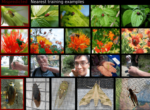
Modern supervised learning algorithms can learn very accurate and complex discriminating functions. But when these classifiers fail, this complexity can also be a drawback because there is no easy, intuitive way to diagnose why they are failing and remedy the problem. This important question has received little attention. To address this problem, we propose a novel method to analyze and understand a classifier's errors. Our method centers around a measure of how much influence a training example has on the classifier's prediction for a test example. To understand why a classifier is mispredicting the label of a given test example, the user can find and review the most influential training examples that caused this misprediction, allowing them to focus their attention on relevant areas of the data space. This will aid the user in determining if and how the training data is inconsistently labeled or lacking in diversity, or if the feature representation is insufficient. As computing the influence of each training example is computationally impractical, we propose a novel distance metric to approximate influence for boosting classifiers that is fast enough to be used interactively. We also show several novel use paradigms of our distance metric. Through experiments, we show that it can be used to find incorrectly or inconsistently labeled training examples, to find specific areas of the data space that need more training data, and to gain insight into which features are missing from the current representation. Code is available at https://github.com/kristinbranson/InfluentialNeighbors.
In practice, understanding the spatial relationships between the surfaces of an object, can significantly improve the performance of object recognition systems. In this paper we propose a novel framework to recognize objects in pictures taken from arbitrary viewpoints. The idea is to maintain the frontal views of the major faces of objects in a global flat map. Then an unfolding warping technique is used to change the pose of the query object in the test view so that all visible surfaces of the object can be observed from a frontal viewpoint, improving the handling of serious occlusions and large viewpoint changes. We demonstrate the effectiveness of our approach through analysis of recognition trials of complex objects with comparison to popular methods.
It is known that sensory deprivation, including postnatal whisker trimming, can lead to severe deficits in the firing rate properties of cortical neurons. Recent results indicate that development of synchronous discharge among cortical neurons is also activity influenced, and that correlated discharge is significantly impaired following loss of bilateral sensory input in rats. Here we investigate whether unilateral whisker trimming (unilateral deprivation or UD) after birth interferes in the same way with the development of synchronous discharge in cortex. We measured the coincidence of spikes among pairs of neurons recorded under urethane anesthesia in one whisker barrel field deprived by trimming all contralateral whiskers for 60 days after birth (UD), and in untrimmed controls (CON). In the septal columns around barrels, UD significantly increased the coincident discharge among cortical neurons compared with CON, most notably in layers II/III. In contrast, synchronous discharge was normal between layer IV UD barrel neurons: i.e., not different from CON. Thus, while bilateral whisker deprivation (BD) produced a global deficit in the development of synchrony in layer IV, UD did not block the development of synchrony between neurons in layer IV barrels and increased synchrony within septal circuits. We conclude that changes in synchronous discharge after UD are unexpectedly different from those recorded after BD, and we speculate that this effect may be due to the driven activity from active commissural inputs arising from the contralateral hemisphere that received normal activity levels during postnatal development.
Gaining independent genetic access to discrete cell types is critical to interrogate their biological functions as well as to deliver precise gene therapy. Transcriptomics has allowed us to profile cell populations with extraordinary precision, revealing that cell types are typically defined by a unique combination of genetic markers. Given the lack of adequate tools to target cell types based on multiple markers, most cell types remain inaccessible to genetic manipulation. Here we present CaSSA, a platform to create unlimited genetic switches based on CRISPR/Cas9 (Ca) and the DNA repair mechanism known as single-strand annealing (SSA). CaSSA allows engineering of independent genetic switches, each responding to a specific gRNA. Expressing multiple gRNAs in specific patterns enables multiplex cell-type-specific manipulations and combinatorial genetic targeting. CaSSA is a new genetic tool that conceptually works as an unlimited number of recombinases and will facilitate genetic access to cell types in diverse organisms.