Filter
Associated Lab
- Aso Lab (8) Apply Aso Lab filter
- Branson Lab (1) Apply Branson Lab filter
- Card Lab (4) Apply Card Lab filter
- Cardona Lab (2) Apply Cardona Lab filter
- Darshan Lab (1) Apply Darshan Lab filter
- Dickson Lab (4) Apply Dickson Lab filter
- Espinosa Medina Lab (2) Apply Espinosa Medina Lab filter
- Fitzgerald Lab (1) Apply Fitzgerald Lab filter
- Funke Lab (1) Apply Funke Lab filter
- Heberlein Lab (1) Apply Heberlein Lab filter
- Hermundstad Lab (2) Apply Hermundstad Lab filter
- Jayaraman Lab (2) Apply Jayaraman Lab filter
- Looger Lab (1) Apply Looger Lab filter
- Reiser Lab (6) Apply Reiser Lab filter
- Romani Lab (2) Apply Romani Lab filter
- Rubin Lab (8) Apply Rubin Lab filter
- Stern Lab (3) Apply Stern Lab filter
- Truman Lab (2) Apply Truman Lab filter
- Turner Lab (4) Apply Turner Lab filter
- Zlatic Lab (1) Apply Zlatic Lab filter
Associated Project Team
Associated Support Team
- Remove Fly Facility filter Fly Facility
- Janelia Experimental Technology (6) Apply Janelia Experimental Technology filter
- Molecular Genomics (3) Apply Molecular Genomics filter
- Project Technical Resources (13) Apply Project Technical Resources filter
- Quantitative Genomics (4) Apply Quantitative Genomics filter
- Scientific Computing Software (3) Apply Scientific Computing Software filter
39 Janelia Publications
Showing 11-20 of 39 resultsWe present CLADES (Cell Lineage Access Driven by an Edition Sequence), a technology for cell lineage studies based on CRISPR/Cas9. CLADES relies on a system of genetic switches to activate and inactivate reporter genes in a pre-determined order. Targeting CLADES to progenitor cells allows the progeny to inherit a sequential cascade of reporters, coupling birth order with reporter expression. This gives us temporal resolution of lineage development that can be used to deconstruct an extended cell lineage by tracking the reporters expressed in the progeny. When targeted to the germ line, the same cascade progresses across animal generations, marking each generation with the corresponding combination of reporters. CLADES thus offers an innovative strategy for making programmable cascades of genes that can be used for genetic manipulation or to record serial biological events.
Wiring a complex brain requires many neurons with intricate cell specificity, generated by a limited number of neural stem cells. central brain lineages are a predetermined series of neurons, born in a specific order. To understand how lineage identity translates to neuron morphology, we mapped 18 central brain lineages. While we found large aggregate differences between lineages, we also discovered shared patterns of morphological diversification. Lineage identity plus Notch-mediated sister fate govern primary neuron trajectories, whereas temporal fate diversifies terminal elaborations. Further, morphological neuron types may arise repeatedly, interspersed with other types. Despite the complexity, related lineages produce similar neuron types in comparable temporal patterns. Different stem cells even yield two identical series of dopaminergic neuron types, but with unrelated sister neurons. Together, these phenomena suggest that straightforward rules drive incredible neuronal complexity, and that large changes in morphology can result from relatively simple fating mechanisms.
The mushroom body (MB) is the center for associative learning in insects. In Drosophila, intersectional split-GAL4 drivers and electron microscopy (EM) connectomes have laid the foundation for precise interrogation of the MB neural circuits. However, many cell types upstream and downstream of the MB remained to be investigated due to lack of driver lines. Here we describe a new collection of over 800 split-GAL4 and split-LexA drivers that cover approximately 300 cell types, including sugar sensory neurons, putative nociceptive ascending neurons, olfactory and thermo-/hygro-sensory projection neurons, interneurons connected with the MB-extrinsic neurons, and various other cell types. We characterized activation phenotypes for a subset of these lines and identified the sugar sensory neuron line most suitable for reward substitution. Leveraging the thousands of confocal microscopy images associated with the collection, we analyzed neuronal morphological stereotypy and discovered that one set of mushroom body output neurons, MBON08/MBON09, exhibits striking individuality and asymmetry across animals. In conjunction with the EM connectome maps, the driver lines reported here offer a powerful resource for functional dissection of neural circuits for associative learning in adult Drosophila.
The nearly constant downward force of gravity has powerfully shaped the behaviors of many organisms [1]. Walking flies readily orient against gravity in a behavior termed negative gravitaxis. In Drosophila this behavior is studied by observing the position of flies in vials [2–4] or simple mazes [5–9]. These assays have been used to conduct forward-genetic screens [5, 6, 8] and as simple tests of locomotion deficits [10–12]. Despite this long history of investigation, the sensory basis of gravitaxis is largely unknown [1]. Recent studies have implicated the antennae as a major mechanosensory input [3, 4], but many details remain unclear. Fly orientation behavior is expected to depend on the direction and amplitude of the gravitational pull, but little is known about the sensitivity of flies to these features of the environment. Here we directly measure the gravity-dependent orientation behavior of flies walking on an adjustable tilted platform, that is inspired by previous insect studies [13–16]. In this arena, flies can freely orient with respect to gravity. Our findings indicate that flies are exquisitely sensitive to the direction of gravity’s pull. Surprisingly, this orientation behavior does not require antennal mechanosensory input, suggesting that other sensory structures must be involved.
Animals perform or terminate particular behaviors by integrating external cues and internal states through neural circuits. Identifying neural substrates and their molecular modulators promoting or inhibiting animal behaviors are key steps to understand how neural circuits control behaviors. Here, we identify the Cholecystokinin-like peptide Drosulfakinin (DSK) that functions at single-neuron resolution to suppress male sexual behavior in Drosophila. We found that Dsk neurons physiologically interact with male-specific P1 neurons, part of a command center for male sexual behaviors, and function oppositely to regulate multiple arousal-related behaviors including sex, sleep and spontaneous walking. We further found that the DSK-2 peptide functions through its receptor CCKLR-17D3 to suppress sexual behaviors in flies. Such a neuropeptide circuit largely overlaps with the fruitless-expressing neural circuit that governs most aspects of male sexual behaviors. Thus DSK/CCKLR signaling in the sex circuitry functions antagonistically with P1 neurons to balance arousal levels and modulate sexual behaviors.
Many animals rely on vision to navigate through their environment. The pattern of changes in the visual scene induced by self-motion is the optic flow1, which is first estimated in local patches by directionally selective (DS) neurons2–4. But how should the arrays of DS neurons, each responsive to motion in a preferred direction at a specific retinal position, be organized to support robust decoding of optic flow by downstream circuits? Understanding this global organization is challenging because it requires mapping fine, local features of neurons across the animal’s field of view3. In Drosophila, the asymmetric dendrites of the T4 and T5 DS neurons establish their preferred direction, making it possible to predict DS responses from anatomy4,5. Here we report that the preferred directions of fly DS neurons vary at different retinal positions and show that this spatial variation is established by the anatomy of the compound eye. To estimate the preferred directions across the visual field, we reconstructed hundreds of T4 neurons in a full brain EM volume6 and discovered unexpectedly stereotypical dendritic arborizations that are independent of location. We then used whole-head μCT scans to map the viewing directions of all compound eye facets and found a non-uniform sampling of visual space that explains the spatial variation in preferred directions. Our findings show that the organization of preferred directions in the fly is largely determined by the compound eye, exposing an intimate and unexpected connection between the peripheral structure of the eye, functional properties of neurons deep in the brain, and the control of body movements.
Internal representations are thought to support the generation of flexible, long-timescale behavioral patterns in both animals and artificial agents. Here, we present a novel conceptual framework for how Drosophila use their internal representation of head direction to maintain preferred headings in their surroundings, and how they learn to modify these preferences in the presence of selective thermal reinforcement. To develop the framework, we analyzed flies’ behavior in a classical operant visual learning paradigm and found that they use stochastically generated fixations and directed turns to express their heading preferences. Symmetries in the visual scene used in the paradigm allowed us to expose how flies’ probabilistic behavior in this setting is tethered to their head direction representation. We describe how flies’ ability to quickly adapt their behavior to the rules of their environment may rest on a behavioral policy whose parameters are flexible but whose form is genetically encoded in the structure of their circuits. Many of the mechanisms we outline may also be relevant for rapidly adaptive behavior driven by internal representations in other animals, including mammals.
Memory guides behavior across widely varying environments and must therefore be both sufficiently specific and general. A memory too specific will be useless in even a slightly different environment, while an overly general memory may lead to suboptimal choices. Animals successfully learn to both distinguish between very similar stimuli and generalize across cues. Rather than forming memories that strike a balance between specificity and generality, Drosophila can flexibly categorize a given stimulus into different groups depending on the options available. We asked how this flexibility manifests itself in the well-characterized learning and memory pathways of the fruit fly. We show that flexible categorization in neuronal activity as well as behavior depends on the order and identity of the perceived stimuli. Our results identify the neural correlates of flexible stimulus-categorization in the fruit fly.
To effectively control their bodies, animals rely on feedback from proprioceptive mechanosensory neurons. In the Drosophila leg, different proprioceptor subtypes monitor joint position, movement direction, and vibration. Here, we investigate how these diverse sensory signals are integrated by central proprioceptive circuits. We find that signals for leg joint position and directional movement converge in second-order neurons, revealing pathways for local feedback control of leg posture. Distinct populations of second-order neurons integrate tibia vibration signals across pairs of legs, suggesting a role in detecting external substrate vibration. In each pathway, the flow of sensory information is dynamically gated and sculpted by inhibition. Overall, our results reveal parallel pathways for processing of internal and external mechanosensory signals, which we propose mediate feedback control of leg movement and vibration sensing, respectively. The existence of a functional connectivity map also provides a resource for interpreting connectomic reconstruction of neural circuits for leg proprioception.
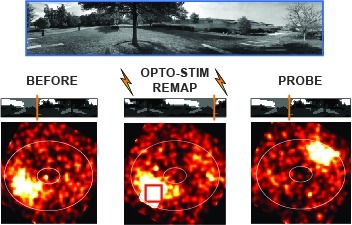
Many animals rely on an internal heading representation when navigating in varied environments. How this representation is linked to the sensory cues that define different surroundings is unclear. In the fly brain, heading is represented by 'compass' neurons that innervate a ring-shaped structure known as the ellipsoid body. Each compass neuron receives inputs from 'ring' neurons that are selective for particular visual features; this combination provides an ideal substrate for the extraction of directional information from a visual scene. Here we combine two-photon calcium imaging and optogenetics in tethered flying flies with circuit modelling, and show how the correlated activity of compass and visual neurons drives plasticity, which flexibly transforms two-dimensional visual cues into a stable heading representation. We also describe how this plasticity enables the fly to convert a partial heading representation, established from orienting within part of a novel setting, into a complete heading representation. Our results provide mechanistic insight into the memory-related computations that are essential for flexible navigation in varied surroundings.