Filter
Associated Lab
- Dickson Lab (1) Apply Dickson Lab filter
- Druckmann Lab (1) Apply Druckmann Lab filter
- Dudman Lab (1) Apply Dudman Lab filter
- Jayaraman Lab (1) Apply Jayaraman Lab filter
- Magee Lab (1) Apply Magee Lab filter
- Menon Lab (1) Apply Menon Lab filter
- Rubin Lab (1) Apply Rubin Lab filter
- Simpson Lab (1) Apply Simpson Lab filter
- Stern Lab (1) Apply Stern Lab filter
- Wu Lab (1) Apply Wu Lab filter
Associated Project Team
Associated Support Team
Publication Date
- February 24, 2014 (1) Apply February 24, 2014 filter
- February 23, 2014 (1) Apply February 23, 2014 filter
- February 19, 2014 (1) Apply February 19, 2014 filter
- February 14, 2014 (1) Apply February 14, 2014 filter
- February 9, 2014 (1) Apply February 9, 2014 filter
- February 6, 2014 (2) Apply February 6, 2014 filter
- February 5, 2014 (1) Apply February 5, 2014 filter
- February 3, 2014 (2) Apply February 3, 2014 filter
- February 1, 2014 (1) Apply February 1, 2014 filter
- Remove February 2014 filter February 2014
- Remove 2014 filter 2014
11 Janelia Publications
Showing 1-10 of 11 resultsDespite the importance of the insect nervous system for functional and developmental neuroscience, descriptions of insect brains have suffered from a lack of uniform nomenclature. Ambiguous definitions of brain regions and fiber bundles have contributed to the variation of names used to describe the same structure. The lack of clearly determined neuropil boundaries has made it difficult to document precise locations of neuronal projections for connectomics study. To address such issues, a consortium of neurobiologists studying arthropod brains, the Insect Brain Name Working Group, has established the present hierarchical nomenclature system, using the brain of Drosophila melanogaster as the reference framework, while taking the brains of other taxa into careful consideration for maximum consistency and expandability. The following summarizes the consortium’s nomenclature system and highlights examples of existing ambiguities and remedies for them. This nomenclature is intended to serve as a standard of reference for the study of the brain of Drosophila and other insects.
We describe a method for fully automated detection of chemical synapses in serial electron microscopy images with highly anisotropic axial and lateral resolution, such as images taken on transmission electron microscopes. Our pipeline starts from classification of the pixels based on 3D pixel features, which is followed by segmentation with an Ising model MRF and another classification step, based on object-level features. Classifiers are learned on sparse user labels; a fully annotated data subvolume is not required for training. The algorithm was validated on a set of 238 synapses in 20 serial 7197×7351 pixel images (4.5×4.5×45 nm resolution) of mouse visual cortex, manually labeled by three independent human annotators and additionally re-verified by an expert neuroscientist. The error rate of the algorithm (12% false negative, 7% false positive detections) is better than state-of-the-art, even though, unlike the state-of-the-art method, our algorithm does not require a prior segmentation of the image volume into cells. The software is based on the ilastik learning and segmentation toolkit and the vigra image processing library and is freely available on our website, along with the test data and gold standard annotations (http://www.ilastik.org/synapse-detection/sstem).
View Publication PageBACKGROUND: Male-specific products of the fruitless (fru) gene control the development and function of neuronal circuits that underlie male-specific behaviors in Drosophila, including courtship. Alternative splicing generates at least three distinct Fru isoforms, each containing a different zinc-finger domain. Here, we examine the expression and function of each of these isoforms. RESULTS: We show that most fru(+) cells express all three isoforms, yet each isoform has a distinct function in the elaboration of sexually dimorphic circuitry and behavior. The strongest impairment in courtship behavior is observed in fru(C) mutants, which fail to copulate, lack sine song, and do not generate courtship song in the absence of visual stimuli. Cellular dimorphisms in the fru circuit are dependent on Fru(C) rather than other single Fru isoforms. Removal of Fru(C) from the neuronal classes vAB3 or aSP4 leads to cell-autonomous feminization of arborizations and loss of courtship in the dark. CONCLUSIONS: These data map specific aspects of courtship behavior to the level of single fru isoforms and fru(+) cell types-an important step toward elucidating the chain of causality from gene to circuit to behavior.
BACKGROUND: High-throughput sequencing is gradually replacing microarrays as the preferred method for studying mRNA expression levels, providing nucleotide resolution and accurately measuring absolute expression levels of almost any transcript, known or novel. However, existing microarray data from clinical, pharmaceutical, and academic settings represent valuable and often underappreciated resources, and methods for assessing and improving the quality of these data are lacking. RESULTS: To quantitatively assess the quality of microarray probes, we directly compare RNA-Seq to Agilent microarrays by processing 231 unique samples from the Allen Human Brain Atlas using RNA-Seq. Both techniques provide highly consistent, highly reproducible gene expression measurements in adult human brain, with RNA-Seq slightly outperforming microarray results overall. We show that RNA-Seq can be used as ground truth to assess the reliability of most microarray probes, remove probes with off-target effects, and scale probe intensities to match the expression levels identified by RNA-Seq. These sequencing scaled microarray intensities (SSMIs) provide more reliable, quantitative estimates of absolute expression levels for many genes when compared with unscaled intensities. Finally, we validate this result in two human cell lines, showing that linear scaling factors can be applied across experiments using the same microarray platform. CONCLUSIONS: Microarrays provide consistent, reproducible gene expression measurements, which are improved using RNA-Seq as ground truth. We expect that our strategy could be used to improve probe quality for many data sets from major existing repositories.
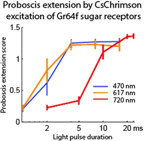
Optogenetic tools enable examination of how specific cell types contribute to brain circuit functions. A long-standing question is whether it is possible to independently activate two distinct neural populations in mammalian brain tissue. Such a capability would enable the study of how different synapses or pathways interact to encode information in the brain. Here we describe two channelrhodopsins, Chronos and Chrimson, discovered through sequencing and physiological characterization of opsins from over 100 species of alga. Chrimson’s excitation spectrum is red shifted by 45 nm relative to previous channelrhodopsins and can enable experiments in which red light is preferred. We show minimal visual system-mediated behavioral interference when using Chrimson in neurobehavioral studies in Drosophila melanogaster. Chronos has faster kinetics than previous channelrhodopsins yet is effectively more light sensitive. Together these two reagents enable two-color activation of neural spiking and downstream synaptic transmission in independent neural populations without detectable cross-talk in mouse brain slice.
The quality of genetically encoded calcium indicators (GECIs) has improved dramatically in recent years, but high-performing ratiometric indicators are still rare. Here we describe a series of fluorescence resonance energy transfer (FRET)-based calcium biosensors with a reduced number of calcium binding sites per sensor. These ’Twitch’ sensors are based on the C-terminal domain of Opsanus troponin C. Their FRET responses were optimized by a large-scale functional screen in bacterial colonies, refined by a secondary screen in rat hippocampal neuron cultures. We tested the in vivo performance of the most sensitive variants in the brain and lymph nodes of mice. The sensitivity of the Twitch sensors matched that of synthetic calcium dyes and allowed visualization of tonic action potential firing in neurons and high resolution functional tracking of T lymphocytes. Given their ratiometric readout, their brightness, large dynamic range and linear response properties, Twitch sensors represent versatile tools for neuroscience and immunology.

A number of recent studies have provided compelling demonstrations that both mice and rats can be trained to perform a variety of behavioral tasks while restrained by mechanical elements mounted to the skull. The independent development of this technique by a number of laboratories has led to diverse solutions. We found that these solutions often used expensive materials and impeded future development and modification in the absence of engineering support. In order to address these issues, here we report on the development of a flexible single hardware design for electrophysiology and imaging both in brain tissue in vitro. Our hardware facilitates the rapid conversion of a single preparation between physiology and imaging system and the conversion of a given system between preparations. In addition, our use of rapid prototyping machines ("3D printers") allows for the deployment of new designs within a day. Here, we present specifications for design and manufacturing as well as some data from our lab demonstrating the suitability of the design for physiology in behaving animals and imaging in vitro and in vivo.
Sparse coding may be a general strategy of neural systems for augmenting memory capacity. In Drosophila melanogaster, sparse odor coding by the Kenyon cells of the mushroom body is thought to generate a large number of precisely addressable locations for the storage of odor-specific memories. However, it remains untested how sparse coding relates to behavioral performance. Here we demonstrate that sparseness is controlled by a negative feedback circuit between Kenyon cells and the GABAergic anterior paired lateral (APL) neuron. Systematic activation and blockade of each leg of this feedback circuit showed that Kenyon cells activated APL and APL inhibited Kenyon cells. Disrupting the Kenyon cell-APL feedback loop decreased the sparseness of Kenyon cell odor responses, increased inter-odor correlations and prevented flies from learning to discriminate similar, but not dissimilar, odors. These results suggest that feedback inhibition suppresses Kenyon cell activity to maintain sparse, decorrelated odor coding and thus the odor specificity of memories.
The organization of synaptic connectivity within a neuronal circuit is a prime determinant of circuit function. We performed a comprehensive fine-scale circuit mapping of hippocampal regions (CA3-CA1) using the newly developed synapse labeling method, mGRASP. This mapping revealed spatially nonuniform and clustered synaptic connectivity patterns. Furthermore, synaptic clustering was enhanced between groups of neurons that shared a similar developmental/migration time window, suggesting a mechanism for establishing the spatial structure of synaptic connectivity. Such connectivity patterns are thought to effectively engage active dendritic processing and storage mechanisms, thereby potentially enhancing neuronal feature selectivity.
Histone variant H2A.Z-containing nucleosomes exist at most eukaryotic promoters and play important roles in gene transcription and genome stability. The multisubunit nucleosome-remodeling enzyme complex SWR1, conserved from yeast to mammals, catalyzes the ATP-dependent replacement of histone H2A in canonical nucleosomes with H2A.Z. How SWR1 catalyzes the replacement reaction is largely unknown. Here, we determined the crystal structure of the N-terminal region (599-627) of the catalytic subunit Swr1, termed Swr1-Z domain, in complex with the H2A.Z-H2B dimer at 1.78 Å resolution. The Swr1-Z domain forms a 310 helix and an irregular chain. A conserved LxxLF motif in the Swr1-Z 310 helix specifically recognizes the αC helix of H2A.Z. Our results show that the Swr1-Z domain can deliver the H2A.Z-H2B dimer to the DNA-(H3-H4)2 tetrasome to form the nucleosome by a histone chaperone mechanism.