Filter
Associated Lab
- Ahrens Lab (1) Apply Ahrens Lab filter
- Remove Aso Lab filter Aso Lab
- Betzig Lab (1) Apply Betzig Lab filter
- Bock Lab (2) Apply Bock Lab filter
- Branson Lab (3) Apply Branson Lab filter
- Cardona Lab (1) Apply Cardona Lab filter
- Clapham Lab (1) Apply Clapham Lab filter
- Funke Lab (2) Apply Funke Lab filter
- Harris Lab (1) Apply Harris Lab filter
- Heberlein Lab (1) Apply Heberlein Lab filter
- Hess Lab (2) Apply Hess Lab filter
- Lippincott-Schwartz Lab (1) Apply Lippincott-Schwartz Lab filter
- Rubin Lab (29) Apply Rubin Lab filter
- Saalfeld Lab (1) Apply Saalfeld Lab filter
- Scheffer Lab (2) Apply Scheffer Lab filter
- Simpson Lab (1) Apply Simpson Lab filter
- Truman Lab (1) Apply Truman Lab filter
- Turner Lab (7) Apply Turner Lab filter
- Zlatic Lab (1) Apply Zlatic Lab filter
Associated Project Team
Associated Support Team
- Electron Microscopy (1) Apply Electron Microscopy filter
- Fly Facility (8) Apply Fly Facility filter
- Project Technical Resources (9) Apply Project Technical Resources filter
- Quantitative Genomics (2) Apply Quantitative Genomics filter
- Scientific Computing Software (3) Apply Scientific Computing Software filter
- Scientific Computing Systems (1) Apply Scientific Computing Systems filter
Publication Date
- 2024 (3) Apply 2024 filter
- 2023 (6) Apply 2023 filter
- 2022 (1) Apply 2022 filter
- 2021 (1) Apply 2021 filter
- 2020 (3) Apply 2020 filter
- 2019 (3) Apply 2019 filter
- 2018 (4) Apply 2018 filter
- 2017 (3) Apply 2017 filter
- 2016 (2) Apply 2016 filter
- 2015 (6) Apply 2015 filter
- 2014 (3) Apply 2014 filter
- 2013 (1) Apply 2013 filter
- 2012 (2) Apply 2012 filter
- 2011 (1) Apply 2011 filter
39 Janelia Publications
Showing 1-10 of 39 resultsUnderstanding memory formation, storage and retrieval requires knowledge of the underlying neuronal circuits. In Drosophila, the mushroom body (MB) is the major site of associative learning. We reconstructed the morphologies and synaptic connections of all 983 neurons within the three functional units, or compartments, that compose the adult MB’s α lobe, using a dataset of isotropic 8-nm voxels collected by focused ion-beam milling scanning electron microscopy. We found that Kenyon cells (KCs), whose sparse activity encodes sensory information, each make multiple en passant synapses to MB output neurons (MBONs) in each compartment. Some MBONs have inputs from all KCs, while others differentially sample sensory modalities. Only six percent of KC>MBON synapses receive a direct synapse from a dopaminergic neuron (DAN). We identified two unanticipated classes of synapses, KC>DAN and DAN>MBON. DAN activation produces a slow depolarization of the MBON in these DAN>MBON synapses and can weaken memory recall.
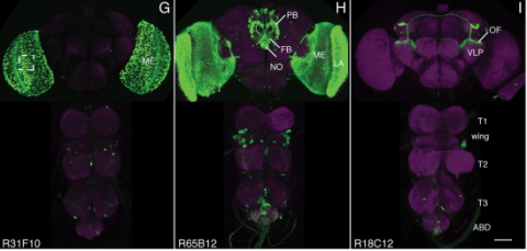
Taste memories allow animals to modulate feeding behavior in accordance with past experience and avoid the consumption of potentially harmful food [1]. We have developed a single-fly taste memory assay to functionally interrogate the neural circuitry encoding taste memories [2]. Here, we screen a collection of Split-GAL4 lines that label small populations of neurons associated with the fly memory center-the mushroom bodies (MBs) [3]. Genetic silencing of PPL1 dopamine neurons disrupts conditioned, but not naive, feeding behavior, suggesting these neurons are selectively involved in the conditioned taste response. We identify two PPL1 subpopulations that innervate the MB α lobe and are essential for aversive taste memory. Thermogenetic activation of these dopamine neurons during training induces memory, indicating these neurons are sufficient for the reinforcing properties of bitter tastant to the MBs. Silencing of either the intrinsic MB neurons or the output neurons from the α lobe disrupts taste conditioning. Thermogenetic manipulation of these output neurons alters naive feeding response, suggesting that dopamine neurons modulate the threshold of response to appetitive tastants. Taken together, these findings detail a neural mechanism underlying the formation of taste memory and provide a functional model for dopamine-dependent plasticity in Drosophila.
We established a collection of 7,000 transgenic lines of Drosophila melanogaster. Expression of GAL4 in each line is controlled by a different, defined fragment of genomic DNA that serves as a transcriptional enhancer. We used confocal microscopy of dissected nervous systems to determine the expression patterns driven by each fragment in the adult brain and ventral nerve cord. We present image data on 6,650 lines. Using both manual and machine-assisted annotation, we describe the expression patterns in the most useful lines. We illustrate the utility of these data for identifying novel neuronal cell types, revealing brain asymmetry, and describing the nature and extent of neuronal shape stereotypy. The GAL4 lines allow expression of exogenous genes in distinct, small subsets of the adult nervous system. The set of DNA fragments, each driving a documented expression pattern, will facilitate the generation of additional constructs for manipulating neuronal function. synapse was substantially elevated, at the endocytic zone there was no enhanced polymerization activity. We conclude that actin subserves spatially diverse, independently regulated processes throughout spines. Perisynaptic actin forms a uniquely dynamic structure well suited for direct, active regulation of the synapse.
For the overall strategy and methods used to produce the GAL4 lines:
Pfeiffer, B.D., Jenett, A., Hammonds, A.S., Ngo, T.T., Misra, S., Murphy, C., Scully, A., Carlson, J.W., Wan, K.H., Laverty, T.R., Mungall, C., Svirskas, R., Kadonaga, J.T., Doe, C.Q., Eisen, M.B., Celniker, S.E., Rubin, G.M. (2008). Tools for neuroanatomy and neurogenetics in Drosophila. Proc. Natl. Acad. Sci. USA 105, 9715-9720. http://www.pnas.org/content/105/28/9715.full.pdf+html synapse was substantially elevated, at the endocytic zone there was no enhanced polymerization activity. We conclude that actin subserves spatially diverse, independently regulated processes throughout spines. Perisynaptic actin forms a uniquely dynamic structure well suited for direct, active regulation of the synapse.
For data on expression in the embryo:
Manning, L., Purice, M.D., Roberts, J., Pollard, J.L., Bennett, A.L., Kroll, J.R., Dyukareva, A.V., Doan, P.N., Lupton, J.R., Strader, M.E., Tanner, S., Bauer, D., Wilbur, A., Tran, K.D., Laverty, T.R., Pearson, J.C., Crews, S.T., Rubin, G.M., and Doe, C.Q. (2012) Annotated embryonic CNS expression patterns of 5000 GMR GAL4 lines: a resource for manipulating gene expression and analyzing cis-regulatory motifs. Cell Reports (2012) Doi: 10.1016/j.celrep.2012.09.009 http://www.cell.com/cell-reports/fulltext/S2211-1247(12)00290-2 synapse was substantially elevated, at the endocytic zone there was no enhanced polymerization activity. We conclude that actin subserves spatially diverse, independently regulated processes throughout spines. Perisynaptic actin forms a uniquely dynamic structure well suited for direct, active regulation of the synapse.
For data on expression in imaginal discs:
Jory, A., Estella, C., Giorgianni, M.W., Slattery, M., Laverty, T.R., Rubin, G.M., and Mann, R.S. (2012) A survey of 6300 genomic fragments for cis-regulatory activity in the imaginal discs of Drosophila melanogaster. Cell Reports (2012) Doi: 10.1016/j.celrep.2012.09.010 http://www.cell.com/cell-reports/fulltext/S2211-1247(12)00291-4 synapse was substantially elevated, at the endocytic zone there was no enhanced polymerization activity. We conclude that actin subserves spatially diverse, independently regulated processes throughout spines. Perisynaptic actin forms a uniquely dynamic structure well suited for direct, active regulation of the synapse.
For data on expression in the larval nervous system:
Li, H.-H., Kroll, J.R., Lennox, S., Ogundeyi, O., Jeter, J., Depasquale, G., and Truman, J.W. (2013) A GAL4 driver resource for developmental and behavioral studies on the larval CNS of Drosophila. Cell Reports (submitted).
Dopamine (DA) is a neurotransmitter with conserved behavioral roles between invertebrate and vertebrate animals. In addition to its neural functions, in insects DA is a critical substrate for cuticle pigmentation and hardening. Drosophila tyrosine hydroxylase (DTH) is the rate limiting enzyme for DA biosynthesis. Viable brain DA deficient flies were previously generated using tissue selective GAL4-UAS binary expression rescue of a DTH null mutation and these flies show specific behavioral impairments. To circumvent the limitations of rescue via binary expression, here we achieve rescue utilizing genomically integrated mutant DTH. As expected, our DA deficient flies have no detectable DTH or DA in the brain, and show reduced locomotor activity. This deficit can be rescued by L-DOPA/carbidopa feeding, similar to human Parkinson's disease treatment. Genetic rescue via GAL4/UAS-DTH was also successful, although this required the generation of a new UAS-DTH1 transgene devoid of most untranslated regions, since existing UAS-DTH transgenes express in the brain without a Gal4 driver via endogenous regulatory elements. A surprising finding of our newly constructed UAS-DTH1m is that it expresses DTH at an undetectable level when regulated by dopaminergic GAL4 drivers even when fully rescuing DA, indicating that DTH immunostaining is not necessarily a valid marker for DA expression. This finding necessitated optimizing DA immunohistochemistry, revealing details of DA innervation to the mushroom body and the central complex. When DA rescue is limited to specific DA neurons, DA does not diffuse beyond the DTH-expressing terminals, such that DA signaling can be limited to very specific brain regions.
Precise, repeatable genetic access to specific neurons via GAL4/UAS and related methods is a key advantage of Drosophila neuroscience. Neuronal targeting is typically documented using light microscopy of full GAL4 expression patterns, which generally lack the single-cell resolution required for reliable cell type identification. Here we use stochastic GAL4 labeling with the MultiColor FlpOut approach to generate cellular resolution confocal images at large scale. We are releasing aligned images of 74,000 such adult central nervous systems. An anticipated use of this resource is to bridge the gap between neurons identified by electron or light microscopy. Identifying individual neurons that make up each GAL4 expression pattern improves the prediction of split-GAL4 combinations targeting particular neurons. To this end we have made the images searchable on the NeuronBridge website. We demonstrate the potential of NeuronBridge to rapidly and effectively identify neuron matches based on morphology across imaging modalities and datasets.
Animals approach stimuli that predict a pleasant outcome. After the paired presentation of an odour and a reward, Drosophila melanogaster can develop a conditioned approach towards that odour. Despite recent advances in understanding the neural circuits for associative memory and appetitive motivation, the cellular mechanisms for reward processing in the fly brain are unknown. Here we show that a group of dopamine neurons in the protocerebral anterior medial (PAM) cluster signals sugar reward by transient activation and inactivation of target neurons in intact behaving flies. These dopamine neurons are selectively required for the reinforcing property of, but not a reflexive response to, the sugar stimulus. In vivo calcium imaging revealed that these neurons are activated by sugar ingestion and the activation is increased on starvation. The output sites of the PAM neurons are mainly localized to the medial lobes of the mushroom bodies (MBs), where appetitive olfactory associative memory is formed. We therefore propose that the PAM cluster neurons endow a positive predictive value to the odour in the MBs. Dopamine in insects is known to mediate aversive reinforcement signals. Our results highlight the cellular specificity underlying the various roles of dopamine and the importance of spatially segregated local circuits within the MBs.
Aggressive social interactions are used to compete for limited resources and are regulated by complex sensory cues and the organism's internal state. While both sexes exhibit aggression, its neuronal underpinnings are understudied in females. Here, we identify a population of sexually dimorphic aIPg neurons in the adult central brain whose optogenetic activation increased, and genetic inactivation reduced, female aggression. Analysis of GAL4 lines identified in an unbiased screen for increased female chasing behavior revealed the involvement of another sexually dimorphic neuron, pC1d, and implicated aIPg and pC1d neurons as core nodes regulating female aggression. Connectomic analysis demonstrated that aIPg neurons and pC1d are interconnected and suggest that aIPg neurons may exert part of their effect by gating the flow of visual information to descending neurons. Our work reveals important regulatory components of the neuronal circuitry that underlies female aggressive social interactions and provides tools for their manipulation.
The behavioral response to a sensory stimulus may depend on both learned and innate neuronal representations. How these circuits interact to produce appropriate behavior is unknown. In Drosophila, the lateral horn (LH) and mushroom body (MB) are thought to mediate innate and learned olfactory behavior, respectively, although LH function has not been tested directly. Here we identify two LH cell types (PD2a1 and PD2b1) that receive input from an MB output neuron required for recall of aversive olfactory memories. These neurons are required for aversive memory retrieval and modulated by training. Connectomics data demonstrate that PD2a1 and PD2b1 neurons also receive direct input from food odor-encoding neurons. Consistent with this, PD2a1 and PD2b1 are also necessary for unlearned attraction to some odors, indicating that these neurons have a dual behavioral role. This provides a circuit mechanism by which learned and innate olfactory information can interact in identified neurons to produce appropriate behavior.
Wiring a complex brain requires many neurons with intricate cell specificity, generated by a limited number of neural stem cells. central brain lineages are a predetermined series of neurons, born in a specific order. To understand how lineage identity translates to neuron morphology, we mapped 18 central brain lineages. While we found large aggregate differences between lineages, we also discovered shared patterns of morphological diversification. Lineage identity plus Notch-mediated sister fate govern primary neuron trajectories, whereas temporal fate diversifies terminal elaborations. Further, morphological neuron types may arise repeatedly, interspersed with other types. Despite the complexity, related lineages produce similar neuron types in comparable temporal patterns. Different stem cells even yield two identical series of dopaminergic neuron types, but with unrelated sister neurons. Together, these phenomena suggest that straightforward rules drive incredible neuronal complexity, and that large changes in morphology can result from relatively simple fating mechanisms.
The Drosophila mushroom body (MB) is an associative learning network that is important for the control of sleep. We have recently identified particular intrinsic MB Kenyon cell (KC) classes that regulate sleep through synaptic activation of particular MB output neurons (MBONs) whose axons convey sleep control signals out of the MB to downstream target regions. Specifically, we found that sleep-promoting KCs increase sleep by preferentially activating cholinergic sleep-promoting MBONs, while wake-promoting KCs decrease sleep by preferentially activating glutamatergic wake-promoting MBONs. Here we use a combination of genetic and physiological approaches to identify wake-promoting dopaminergic neurons (DANs) that innervate the MB, and show that they activate wake-promoting MBONs. These studies reveal a dopaminergic sleep control mechanism that likely operates by modulation of KC-MBON microcircuits.