Filter
Associated Lab
- Betzig Lab (2) Apply Betzig Lab filter
- Eddy/Rivas Lab (2) Apply Eddy/Rivas Lab filter
- Hess Lab (1) Apply Hess Lab filter
- Karpova Lab (1) Apply Karpova Lab filter
- Lippincott-Schwartz Lab (1) Apply Lippincott-Schwartz Lab filter
- Rubin Lab (2) Apply Rubin Lab filter
- Shroff Lab (1) Apply Shroff Lab filter
- Spruston Lab (4) Apply Spruston Lab filter
- Svoboda Lab (3) Apply Svoboda Lab filter
- Tervo Lab (1) Apply Tervo Lab filter
Publication Date
- December 2007 (5) Apply December 2007 filter
- November 2007 (1) Apply November 2007 filter
- October 2007 (2) Apply October 2007 filter
- July 2007 (3) Apply July 2007 filter
- June 2007 (1) Apply June 2007 filter
- May 2007 (3) Apply May 2007 filter
- April 2007 (3) Apply April 2007 filter
- March 2007 (2) Apply March 2007 filter
- February 2007 (1) Apply February 2007 filter
- Remove 2007 filter 2007
21 Janelia Publications
Showing 1-10 of 21 resultsStaining the mRNA of a gene via in situ hybridization (ISH) during the development of a D. melanogaster embryo delivers the detailed spatio-temporal pattern of expression of the gene. Many biological problems such as the detection of co-expressed genes, co-regulated genes, and transcription factor binding motifs rely heavily on the analyses of these image patterns. The increasing availability of ISH image data motivates the development of automated computational approaches to the analysis of gene expression patterns.
Gene expression patterns obtained by in situ mRNA hybridization provide important information about different genes during Drosophila embryogenesis. So far, annotations of these images are done by manually assigning a subset of anatomy ontology terms to an image. This time-consuming process depends heavily on the consistency of experts.
Automatic segmentation of nuclei in 3D microscopy images is essential for many biological studies including high throughput analysis of gene expression level, morphology, and phenotypes in single cell level. The complexity and variability of the microscopy images present many difficulties to the traditional image segmentation methods. In this paper, we present a new method based on 3D watershed algorithm to segment such images. By using both the intensity information of the image and the geometry information of the appropriately detected foreground mask, our method is robust to intensity fluctuation within nuclei and at the same time sensitive to the intensity and geometrical cues between nuclei. Besides, the method can automatically correct potential segmentation errors by using several post-processing steps. We tested this algorithm on the 3D confocal images of C.elegans, an organism that has been widely used in biological studies. Our results show that the algorithm can segment nuclei in high accuracy despite the non-uniform background, tightly clustered nuclei with different sizes and shapes, fluctuated intensities, and hollow-shaped staining patterns in the images.
The functions of cortical areas depend on their inputs and outputs, but the detailed circuits made by long-range projections are unknown. We show that the light-gated channel channelrhodopsin-2 (ChR2) is delivered to axons in pyramidal neurons in vivo. In brain slices from ChR2-expressing mice, photostimulation of ChR2-positive axons can be transduced reliably into single action potentials. Combining photostimulation with whole-cell recordings of synaptic currents makes it possible to map circuits between presynaptic neurons, defined by ChR2 expression, and postsynaptic neurons, defined by targeted patching. We applied this technique, ChR2-assisted circuit mapping (CRACM), to map long-range callosal projections from layer (L) 2/3 of the somatosensory cortex. L2/3 axons connect with neurons in L5, L2/3 and L6, but not L4, in both ipsilateral and contralateral cortex. In both hemispheres the L2/3-to-L5 projection is stronger than the L2/3-to-L2/3 projection. Our results suggest that laminar specificity may be identical for local and long-range cortical projections.
Recent advances in single-neuron biophysics have enhanced our understanding of information processing on the cellular level, but how the detailed properties of individual neurons give rise to large-scale behavior remains unclear. Here, we present a model of the hippocampal network based on observed biophysical properties of hippocampal and entorhinal cortical neurons. We assembled our model to simulate spatial alternation, a task that requires memory of the previous path through the environment for correct selection of the current path to a reward site. The convergence of inputs from entorhinal cortex and hippocampal region CA3 onto CA1 pyramidal cells make them potentially important for integrating information about place and temporal context on the network level. Our model shows how place and temporal context information might be combined in CA1 pyramidal neurons to give rise to splitter cells, which fire selectively based on a combination of place and temporal context. The model leads to a number of experimentally testable predictions that may lead to a better understanding of the biophysical basis of information processing in the hippocampus.
This paper presents a new study on a method of designing a multi-class classifier: Data-driven Error Correcting Output Coding (DECOC). DECOC is based on the principle of Error Correcting Output Coding (ECOC), which uses a code matrix to decompose a multi-class problem into multiple binary problems. ECOC for multi-class classification hinges on the design of the code matrix. We propose to explore the distribution of data classes and optimize both the composition and the number of base learners to design an effective and compact code matrix. Two real world applications are studied: (1) the holistic recognition (i.e., recognition without segmentation) of touching handwritten numeral pairs and (2) the classification of cancer tissue types based on microarray gene expression data. The results show that the proposed DECOC is able to deliver competitive accuracy compared with other ECOC methods, using parsimonious base learners than the pairwise coupling (one-vs-one) decomposition scheme. With a rejection scheme defined by a simple robustness measure, high reliabilities of around 98% are achieved in both applications.
In CA1 pyramidal neurons, burst firing is correlated with hippocampally dependent behaviours and modulation of synaptic strength. One of the mechanisms underlying burst firing in these cells is the afterdepolarization (ADP) that follows each action potential. Previous work has shown that the ADP results from the interaction of several depolarizing and hyperpolarizing conductances located in the soma and the dendrites. By using patch-clamp recordings from acute rat hippocampal slices we show that D-type potassium current modulates the size of the ADP and the bursting of CA1 pyramidal neurons. Sensitivity to alpha-dendrotoxin suggests that Kv1-containing potassium channels mediate this current. Dual somato-dendritic recording, outside-out dendritic recordings, and focal application of dendrotoxin together indicate that the channels mediating this current are located in the apical dendrites. Thus, our data present evidence for a dendritic segregation of Kv1-like channels in CA1 pyramidal neurons and identify a novel action for these channels, showing that they inhibit action potential bursting by restricting the size of the ADP.
The hippocampus is essential for episodic memory, which requires single-trial learning. Although long-term potentiation (LTP) of synaptic strength is a candidate mechanism for learning, it is typically induced by using repeated synaptic activation to produce precisely timed, high-frequency, or rhythmic firing. Here we show that hippocampal synapses potentiate robustly in response to strong activation by a single burst. The induction mechanism of this single-burst LTP requires activation of NMDA receptors, L-type voltage-gated calcium channels, and dendritic spikes. Thus, dendritic spikes are a critical trigger for a form of LTP that is consistent with the function of the hippocampus in episodic memory.
In conventional biological imaging, diffraction places a limit on the minimal xy distance at which two marked objects can be discerned. Consequently, resolution of target molecules within cells is typically coarser by two orders of magnitude than the molecular scale at which the proteins are spatially distributed. Photoactivated localization microscopy (PALM) optically resolves selected subsets of protect fluorescent probes within cells at mean separations of <25 nanometers. It involves serial photoactivation and subsequent photobleaching of numerous sparse subsets of photoactivated fluorescent protein molecules. Individual molecules are localized at near molecular resolution by determining their centers of fluorescent emission via a statistical fit of their point-spread-function. The position information from all subsets is then assembled into a super-resolution image, in which individual fluorescent molecules are isolated at high molecular densities. In this paper, some of the limitations for PALM imaging under current experimental conditions are discussed.
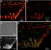
Accurate determination of the relative positions of proteins within localized regions of the cell is essential for understanding their biological function. Although fluorescent fusion proteins are targeted with molecular precision, the position of these genetically expressed reporters is usually known only to the resolution of conventional optics ( approximately 200 nm). Here, we report the use of two-color photoactivated localization microscopy (PALM) to determine the ultrastructural relationship between different proteins fused to spectrally distinct photoactivatable fluorescent proteins (PA-FPs). The nonperturbative incorporation of these endogenous tags facilitates an imaging resolution in whole, fixed cells of approximately 20-30 nm at acquisition times of 5-30 min. We apply the technique to image different pairs of proteins assembled in adhesion complexes, the central attachment points between the cytoskeleton and the substrate in migrating cells. For several pairs, we find that proteins that seem colocalized when viewed by conventional optics are resolved as distinct interlocking nano-aggregates when imaged via PALM. The simplicity, minimal invasiveness, resolution, and speed of the technique all suggest its potential to directly visualize molecular interactions within cellular structures at the nanometer scale.
Commentary: Identifies the photoactivatable fluorescent proteins (PA-FPs) Dronpa and PS-CFP2 as green partners to orange-red PA-FPs such as Kaede and Eos for dual color PALM imaging. Very low crosstalk is demonstrated between the two color channels. Furthermore, since the probes are genetically expressed, they are closely bound to their target proteins and exhibit zero non-specific background. All these properties are essential to unambiguously identify regions of co-localization or separate compartmentalization at the nanoscale, as demonstrated in the examples here.