Filter
Associated Lab
- Ahrens Lab (4) Apply Ahrens Lab filter
- Aso Lab (3) Apply Aso Lab filter
- Betzig Lab (4) Apply Betzig Lab filter
- Beyene Lab (1) Apply Beyene Lab filter
- Branson Lab (3) Apply Branson Lab filter
- Card Lab (5) Apply Card Lab filter
- Cardona Lab (3) Apply Cardona Lab filter
- Clapham Lab (1) Apply Clapham Lab filter
- Dickson Lab (4) Apply Dickson Lab filter
- Dudman Lab (2) Apply Dudman Lab filter
- Espinosa Medina Lab (2) Apply Espinosa Medina Lab filter
- Fitzgerald Lab (3) Apply Fitzgerald Lab filter
- Funke Lab (4) Apply Funke Lab filter
- Grigorieff Lab (3) Apply Grigorieff Lab filter
- Harris Lab (1) Apply Harris Lab filter
- Heberlein Lab (2) Apply Heberlein Lab filter
- Hermundstad Lab (2) Apply Hermundstad Lab filter
- Hess Lab (5) Apply Hess Lab filter
- Jayaraman Lab (4) Apply Jayaraman Lab filter
- Keller Lab (5) Apply Keller Lab filter
- Lavis Lab (9) Apply Lavis Lab filter
- Lee (Albert) Lab (5) Apply Lee (Albert) Lab filter
- Lippincott-Schwartz Lab (8) Apply Lippincott-Schwartz Lab filter
- Liu (Zhe) Lab (7) Apply Liu (Zhe) Lab filter
- Looger Lab (7) Apply Looger Lab filter
- Pachitariu Lab (2) Apply Pachitariu Lab filter
- Podgorski Lab (5) Apply Podgorski Lab filter
- Reiser Lab (2) Apply Reiser Lab filter
- Romani Lab (2) Apply Romani Lab filter
- Rubin Lab (9) Apply Rubin Lab filter
- Saalfeld Lab (2) Apply Saalfeld Lab filter
- Scheffer Lab (1) Apply Scheffer Lab filter
- Schreiter Lab (5) Apply Schreiter Lab filter
- Spruston Lab (5) Apply Spruston Lab filter
- Stern Lab (4) Apply Stern Lab filter
- Sternson Lab (4) Apply Sternson Lab filter
- Stringer Lab (2) Apply Stringer Lab filter
- Svoboda Lab (5) Apply Svoboda Lab filter
- Truman Lab (3) Apply Truman Lab filter
- Turaga Lab (1) Apply Turaga Lab filter
- Turner Lab (3) Apply Turner Lab filter
- Zlatic Lab (2) Apply Zlatic Lab filter
Associated Project Team
- Fly Descending Interneuron (2) Apply Fly Descending Interneuron filter
- Fly Functional Connectome (1) Apply Fly Functional Connectome filter
- FlyEM (3) Apply FlyEM filter
- FlyLight (8) Apply FlyLight filter
- GENIE (5) Apply GENIE filter
- MouseLight (1) Apply MouseLight filter
- Tool Translation Team (T3) (3) Apply Tool Translation Team (T3) filter
- Transcription Imaging (1) Apply Transcription Imaging filter
Associated Support Team
- Anatomy and Histology (3) Apply Anatomy and Histology filter
- Cryo-Electron Microscopy (2) Apply Cryo-Electron Microscopy filter
- Electron Microscopy (1) Apply Electron Microscopy filter
- Fly Facility (10) Apply Fly Facility filter
- Integrative Imaging (2) Apply Integrative Imaging filter
- Janelia Experimental Technology (8) Apply Janelia Experimental Technology filter
- Molecular Genomics (4) Apply Molecular Genomics filter
- Primary & iPS Cell Culture (5) Apply Primary & iPS Cell Culture filter
- Project Technical Resources (3) Apply Project Technical Resources filter
- Quantitative Genomics (3) Apply Quantitative Genomics filter
- Scientific Computing Software (2) Apply Scientific Computing Software filter
- Scientific Computing Systems (2) Apply Scientific Computing Systems filter
- Viral Tools (2) Apply Viral Tools filter
- Vivarium (1) Apply Vivarium filter
Publication Date
- Remove 2020 filter 2020
178 Janelia Publications
Showing 41-50 of 178 results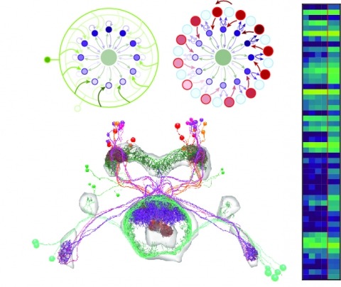
Neural representations of head direction (HD) have been discovered in many species. Theoretical work has proposed that the dynamics associated with these representations are generated, maintained, and updated by recurrent network structures called ring attractors. We evaluated this theorized structure-function relationship by performing electron-microscopy-based circuit reconstruction and RNA profiling of identified cell types in the HD system of Drosophila melanogaster. We identified motifs that have been hypothesized to maintain the HD representation in darkness, update it when the animal turns, and tether it to visual cues. Functional studies provided support for the proposed roles of individual excitatory or inhibitory circuit elements in shaping activity. We also discovered recurrent connections between neuronal arbors with mixed pre- and postsynaptic specializations. Our results confirm that the Drosophila HD network contains the core components of a ring attractor while also revealing unpredicted structural features that might enhance the network's computational power.

An important question in early neural development is the origin of stochastic nuclear movement between apical and basal surfaces of neuroepithelia during interkinetic nuclear migration. Tracking of nuclear subpopulations has shown evidence of diffusion - mean squared displacements growing linearly in time - and suggested crowding from cell division at the apical surface drives basalward motion. Yet, this hypothesis has not yet been tested, and the forces involved not quantified. We employ long-term, rapid light-sheet and two-photon imaging of early zebrafish retinogenesis to track entire populations of nuclei within the tissue. The time-varying concentration profiles show clear evidence of crowding as nuclei reach close-packing and are quantitatively described by a nonlinear diffusion model. Considerations of nuclear motion constrained inside the enveloping cell membrane show that concentration-dependent stochastic forces inside cells, compatible in magnitude to those found in cytoskeletal transport, can explain the observed magnitude of the diffusion constant.
mTOR is a serine/threonine kinase and a master regulator of cell growth and proliferation. Raptor, a scaffolding protein that recruits substrates to mTOR complex 1 (mTORC1), is known to be phosphorylated during mitosis, but the significance of this phosphorylation remains largely unknown. Here we show that raptor expression and mTORC1 activity are dramatically reduced in cells arrested in mitosis. Expression of a non-phosphorylatable raptor mutant reactivates mTORC1 and significantly reduces cytotoxicity of the mitotic poison Taxol. This effect is mediated via degradation of PDCD4, a tumor suppressor protein that inhibits eIF4A activity and is negatively regulated by the mTORC1/S6K pathway. Moreover, pharmacological inhibition of eIF4A is able to enhance the effects of Taxol and restore sensitivity in Taxol-resistant cancer cells. These findings indicate that the mTORC1/S6K/PDCD4/eIF4A axis has a pivotal role in the death versus slippage decision during mitotic arrest and may be exploited clinically to treat tumors resistant to anti-mitotic agents.
The mating decisions of Drosophila melanogaster females are primarily revealed through either of two discrete actions: opening of the vaginal plates to allow copulation, or extrusion of the ovipositor to reject the male. Both actions are triggered by the male courtship song, and both are dependent upon the female's mating status. Virgin females are more likely to open their vaginal plates in response to song; mated females are more likely to extrude their ovipositor. Here, we examine the neural cause and behavioral consequence of ovipositor extrusion. We show that the DNp13 descending neurons act as command-type neurons for ovipositor extrusion, and that ovipositor extrusion is an effective deterrent only when performed by females that have previously mated. The DNp13 neurons respond to male song via direct synaptic input from the pC2l auditory neurons. Mating status does not modulate the song responses of DNp13 neurons, but rather how effectively they can engage the motor circuits for ovipositor extrusion. We present evidence that mating status information is mediated by ppk sensory neurons in the uterus, which are activated upon ovulation. Vaginal plate opening and ovipositor extrusion are thus controlled by anatomically and functionally distinct circuits, highlighting the diversity of neural decision-making circuits even in the context of closely related behaviors with shared exteroceptive and interoceptive inputs.
The striosome compartment within the dorsal striatum has been implicated in reinforcement learning and regulation of motivation, but how striosomal neurons contribute to these functions remains elusive. Here, we show that a genetically identified striosomal population, which expresses the Teashirt family zinc finger 1 (Tshz1) and belongs to the direct pathway, drives negative reinforcement and is essential for aversive learning in mice. Contrasting a "conventional" striosomal direct pathway, the Tshz1 neurons cause aversion, movement suppression, and negative reinforcement once activated, and they receive a distinct set of synaptic inputs. These neurons are predominantly excited by punishment rather than reward and represent the anticipation of punishment or the motivation for avoidance. Furthermore, inhibiting these neurons impairs punishment-based learning without affecting reward learning or movement. These results establish a major role of striosomal neurons in behaviors reinforced by punishment and moreover uncover functions of the direct pathway unaccounted for in classic models.
is a major human pathogen that has acquired alarming broad-spectrum antibiotic resistance. One group of secreted toxins with key roles during infection is the phenol-soluble modulins (PSMs). PSMs are amphipathic, membrane-destructive cytolytic peptides that are exported to the host-cell environment by a designated adenosine 5'-triphosphate (ATP)-binding cassette (ABC) transporter, the PSM transporter (PmtABCD). Here, we demonstrate that the minimal Pmt unit necessary for PSM export is PmtCD and provide its first atomic characterization by single-particle cryo-EM and x-ray crystallography. We have captured the transporter in the ATP-bound state at near atomic resolution, revealing a type II ABC exporter fold, with an additional cytosolic domain. Comparison to a lower-resolution nucleotide-free map displaying an "open" conformation and putative hydrophobic inner chamber of a size able to accommodate the binding of two PSM peptides provides mechanistic insight and sets the foundation for therapeutic design.
Three-dimensional (3D) chromatin organization plays a key role in regulating mammalian genome function; however, many of its physical features at the single-cell level remain underexplored. Here, we use live- and fixed-cell 3D super-resolution and scanning electron microscopy to analyze structural and functional nuclear organization in somatic cells. We identify chains of interlinked ~200- to 300-nm-wide chromatin domains (CDs) composed of aggregated nucleosomes that can overlap with individual topologically associating domains and are distinct from a surrounding RNA-populated interchromatin compartment. High-content mapping uncovers confinement of cohesin and active histone modifications to surfaces and enrichment of repressive modifications toward the core of CDs in both hetero- and euchromatic regions. This nanoscale functional topography is temporarily relaxed in postreplicative chromatin but remarkably persists after ablation of cohesin. Our findings establish CDs as physical and functional modules of mesoscale genome organization.