Filter
Associated Lab
- Remove Ahrens Lab filter Ahrens Lab
- Aso Lab (1) Apply Aso Lab filter
- Branson Lab (1) Apply Branson Lab filter
- Fitzgerald Lab (1) Apply Fitzgerald Lab filter
- Freeman Lab (5) Apply Freeman Lab filter
- Harris Lab (2) Apply Harris Lab filter
- Jayaraman Lab (2) Apply Jayaraman Lab filter
- Johnson Lab (1) Apply Johnson Lab filter
- Keller Lab (5) Apply Keller Lab filter
- Lavis Lab (1) Apply Lavis Lab filter
- Liu (Zhe) Lab (1) Apply Liu (Zhe) Lab filter
- Looger Lab (7) Apply Looger Lab filter
- Podgorski Lab (3) Apply Podgorski Lab filter
- Schreiter Lab (4) Apply Schreiter Lab filter
- Svoboda Lab (4) Apply Svoboda Lab filter
- Turaga Lab (2) Apply Turaga Lab filter
- Turner Lab (2) Apply Turner Lab filter
- Zlatic Lab (1) Apply Zlatic Lab filter
Associated Project Team
Associated Support Team
Publication Date
- 2024 (2) Apply 2024 filter
- 2023 (4) Apply 2023 filter
- 2022 (4) Apply 2022 filter
- 2021 (2) Apply 2021 filter
- 2020 (4) Apply 2020 filter
- 2019 (5) Apply 2019 filter
- 2018 (4) Apply 2018 filter
- 2017 (2) Apply 2017 filter
- 2016 (7) Apply 2016 filter
- 2015 (3) Apply 2015 filter
- 2014 (3) Apply 2014 filter
- 2013 (2) Apply 2013 filter
42 Janelia Publications
Showing 31-40 of 42 resultsIn the absence of salient sensory cues to guide behavior, animals must still execute sequences of motor actions in order to forage and explore. How such successive motor actions are coordinated to form global locomotion trajectories is unknown. We mapped the structure of larval zebrafish swim trajectories in homogeneous environments and found that trajectories were characterized by alternating sequences of repeated turns to the left and to the right. Using whole-brain light-sheet imaging, we identified activity relating to the behavior in specific neural populations that we termed the anterior rhombencephalic turning region (ARTR). ARTR perturbations biased swim direction and reduced the dependence of turn direction on turn history, indicating that the ARTR is part of a network generating the temporal correlations in turn direction. We also find suggestive evidence for ARTR mutual inhibition and ARTR projections to premotor neurons. Finally, simulations suggest the observed turn sequences may underlie efficient exploration of local environments.
Increasing the volumetric imaging speed of light-sheet microscopy will improve its ability to detect fast changes in neural activity. Here, a system is introduced for brain-wide imaging of neural activity in the larval zebrafish by coupling structured illumination with cubic phase extended depth-of-field (EDoF) pupil encoding. This microscope enables faster light-sheet imaging and facilitates arbitrary plane scanning—removing constraints on acquisition speed, alignment tolerances, and physical motion near the sample. The usefulness of this method is demonstrated by performing multi-plane calcium imaging in the fish brain with a 416×832×160 μm field of view at 33 Hz. The optomotor response behavior of the zebrafish is monitored at high speeds, and time-locked correlations of neuronal activity are resolved across its brain.
Escape behaviors deliver organisms away from imminent catastrophe. Here, we characterize behavioral responses of freely swimming larval zebrafish to looming visual stimuli simulating predators. We report that the visual system alone can recruit lateralized, rapid escape motor programs, similar to those elicited by mechanosensory modalities. Two-photon calcium imaging of retino-recipient midbrain regions isolated the optic tectum as an important center processing looming stimuli, with ensemble activity encoding the critical image size determining escape latency. Furthermore, we describe activity in retinal ganglion cell terminals and superficial inhibitory interneurons in the tectum during looming and propose a model for how temporal dynamics in tectal periventricular neurons might arise from computations between these two fundamental constituents. Finally, laser ablations of hindbrain circuitry confirmed that visual and mechanosensory modalities share the same premotor output network. We establish a circuit for the processing of aversive stimuli in the context of an innate visual behavior.
We present a modular approach for analyzing calcium imaging recordings of large neuronal ensembles. Our goal is to simultaneously identify the locations of the neurons, demix spatially overlapping components, and denoise and deconvolve the spiking activity from the slow dynamics of the calcium indicator. Our approach relies on a constrained nonnegative matrix factorization that expresses the spatiotemporal fluorescence activity as the product of a spatial matrix that encodes the spatial footprint of each neuron in the optical field and a temporal matrix that characterizes the calcium concentration of each neuron over time. This framework is combined with a novel constrained deconvolution approach that extracts estimates of neural activity from fluorescence traces, to create a spatiotemporal processing algorithm that requires minimal parameter tuning. We demonstrate the general applicability of our method by applying it to in vitro and in vivo multi-neuronal imaging data, whole-brain light-sheet imaging data, and dendritic imaging data.
The dense connectivity in the brain means that one neuron's activity can influence many others. To observe this interconnected system comprehensively, an aspiration within neuroscience is to record from as many neurons as possible at the same time. There are two useful routes toward this goal: one is to expand the spatial extent of functional imaging techniques, and the second is to use animals with small brains. Here we review recent progress toward imaging many neurons and complete populations of identified neurons in small vertebrates and invertebrates.
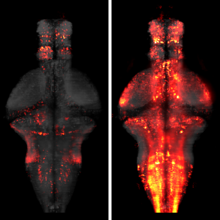
The identification of active neurons and circuits in vivo is a fundamental challenge in understanding the neural basis of behavior. Genetically encoded calcium (Ca(2+)) indicators (GECIs) enable quantitative monitoring of cellular-resolution activity during behavior. However, such indicators require online monitoring within a limited field of view. Alternatively, post hoc staining of immediate early genes (IEGs) indicates highly active cells within the entire brain, albeit with poor temporal resolution. We designed a fluorescent sensor, CaMPARI, that combines the genetic targetability and quantitative link to neural activity of GECIs with the permanent, large-scale labeling of IEGs, allowing a temporally precise "activity snapshot" of a large tissue volume. CaMPARI undergoes efficient and irreversible green-to-red conversion only when elevated intracellular Ca(2+) and experimenter-controlled illumination coincide. We demonstrate the utility of CaMPARI in freely moving larvae of zebrafish and flies, and in head-fixed mice and adult flies.

The nature of nervous system function and development is inherently global, since all components eventually influence one another. Networks communicate through dense synaptic, electric, and modulatory connections and develop through concurrent growth and interlinking of their neurons, processes, glia, and blood vessels. These factors drive the development of techniques capable of imaging neural signaling, anatomy, and developmental processes at ever-larger scales. Here, we discuss the nature of questions benefitting from large-scale imaging techniques and introduce recent applications. We focus on emerging light-sheet microscopy approaches, which are well suited for live imaging of large systems with high spatiotemporal resolution and over long periods of time. We also discuss computational methods suitable for extracting biological information from the resulting system-level image data sets. Together with new tools for reporting and manipulating neuronal activity and gene expression, these techniques promise new insights into the large-scale function and development of neural systems.

Developments in electrical and optical recording technology are scaling up the size of neuronal populations that can be monitored simultaneously. Light-sheet imaging is rapidly gaining traction as a method for optically interrogating activity in large networks and presents both opportunities and challenges for understanding circuit function.
The processing of sensory input and the generation of behavior involves large networks of neurons, which necessitates new technology for recording from many neurons in behaving animals. In the larval zebrafish, light-sheet microscopy can be used to record the activity of almost all neurons in the brain simultaneously at single-cell resolution. Existing implementations, however, cannot be combined with visually driven behavior because the light sheet scans over the eye, interfering with presentation of controlled visual stimuli. Here we describe a system that overcomes the confounding eye stimulation through the use of two light sheets and combines whole-brain light-sheet imaging with virtual reality for fictively behaving larval zebrafish.
Understanding brain function requires monitoring and interpreting the activity of large networks of neurons during behavior. Advances in recording technology are greatly increasing the size and complexity of neural data. Analyzing such data will pose a fundamental bottleneck for neuroscience. We present a library of analytical tools called Thunder built on the open-source Apache Spark platform for large-scale distributed computing. The library implements a variety of univariate and multivariate analyses with a modular, extendable structure well-suited to interactive exploration and analysis development. We demonstrate how these analyses find structure in large-scale neural data, including whole-brain light-sheet imaging data from fictively behaving larval zebrafish, and two-photon imaging data from behaving mouse. The analyses relate neuronal responses to sensory input and behavior, run in minutes or less and can be used on a private cluster or in the cloud. Our open-source framework thus holds promise for turning brain activity mapping efforts into biological insights.