Filter
Associated Lab
- Ahrens Lab (5) Apply Ahrens Lab filter
- Branson Lab (3) Apply Branson Lab filter
- Card Lab (1) Apply Card Lab filter
- Cardona Lab (1) Apply Cardona Lab filter
- Druckmann Lab (1) Apply Druckmann Lab filter
- Freeman Lab (4) Apply Freeman Lab filter
- Funke Lab (2) Apply Funke Lab filter
- Harris Lab (1) Apply Harris Lab filter
- Ji Lab (1) Apply Ji Lab filter
- Remove Keller Lab filter Keller Lab
- Lavis Lab (2) Apply Lavis Lab filter
- Liu (Zhe) Lab (1) Apply Liu (Zhe) Lab filter
- Looger Lab (5) Apply Looger Lab filter
- Pavlopoulos Lab (1) Apply Pavlopoulos Lab filter
- Tillberg Lab (1) Apply Tillberg Lab filter
- Turaga Lab (1) Apply Turaga Lab filter
Associated Project Team
Publication Date
- 2024 (1) Apply 2024 filter
- 2023 (1) Apply 2023 filter
- 2022 (1) Apply 2022 filter
- 2021 (2) Apply 2021 filter
- 2020 (5) Apply 2020 filter
- 2019 (6) Apply 2019 filter
- 2018 (6) Apply 2018 filter
- 2017 (2) Apply 2017 filter
- 2016 (6) Apply 2016 filter
- 2015 (8) Apply 2015 filter
- 2014 (8) Apply 2014 filter
- 2013 (10) Apply 2013 filter
- 2012 (3) Apply 2012 filter
- 2011 (4) Apply 2011 filter
- 2010 (4) Apply 2010 filter
- 2009 (1) Apply 2009 filter
- 2008 (3) Apply 2008 filter
- 2007 (1) Apply 2007 filter
- 2006 (2) Apply 2006 filter
- 2005 (1) Apply 2005 filter
Type of Publication
75 Publications
Showing 51-60 of 75 results
The observation of biological processes in their natural in vivo context is a key requirement for quantitative experimental studies in the life sciences. In many instances, it will be crucial to achieve high temporal and spatial resolution over long periods of time without compromising the physiological development of the specimen. Here, we discuss the principles underlying light sheet-based fluorescence microscopes. The most recent implementation DSLM is a tool optimized to deliver quantitative data for entire embryos at high spatio-temporal resolution. We compare DSLM to the two established light microscopy techniques: confocal and two-photon fluorescence microscopy. DSLM provides up to 50 times higher imaging speeds and a 10-100 times higher signal-to-noise ratio, while exposing the specimens to at least three orders of magnitude less light energy than confocal and two-photon fluorescence microscopes. We conclude with a perspective for future development.

We present the Real-time Accurate Cell-shape Extractor (RACE), a high-throughput image analysis framework for automated three-dimensional cell segmentation in large-scale images. RACE is 55–330 times faster and 2–5 times more accurate than state-of-the-art methods. We demonstrate the generality of RACE by extracting cell-shape information from entire Drosophila, zebrafish, and mouse embryos imaged with confocal and light-sheet microscopes. Using RACE, we automatically reconstructed cellular-resolution tissue anisotropy maps across developing Drosophila embryos and quantified differences in cell-shape dynamics in wild-type and mutant embryos. We furthermore integrated RACE with our framework for automated cell lineaging and performed joint segmentation and cell tracking in entire Drosophila embryos. RACE processed these terabyte-sized datasets on a single computer within 1.4 days. RACE is easy to use, as it requires adjustment of only three parameters, takes full advantage of state-of-the-art multi-core processors and graphics cards, and is available as open-source software for Windows, Linux, and Mac OS.
Novel approaches to bio-imaging and automated computational image processing allow the design of truly quantitative studies in developmental biology. Cell behavior, cell fate decisions, cell interactions during tissue morphogenesis, and gene expression dynamics can be analyzed in vivo for entire complex organisms and throughout embryonic development. We review state-of-the-art technology for live imaging, focusing on fluorescence light microscopy techniques for system-level investigations of animal development and discuss computational approaches to image segmentation, cell tracking, automated data annotation, and biophysical modeling. We argue that the substantial increase in data complexity and size requires sophisticated new strategies to data analysis to exploit the enormous potential of these new resources.
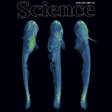
A long-standing goal of biology is to map the behavior of all cells during vertebrate embryogenesis. We developed digital scanned laser light sheet fluorescence microscopy and recorded nuclei localization and movement in entire wild-type and mutant zebrafish embryos over the first 24 hours of development. Multiview in vivo imaging at 1.5 billion voxels per minute provides "digital embryos," that is, comprehensive databases of cell positions, divisions, and migratory tracks. Our analysis of global cell division patterns reveals a maternally defined initial morphodynamic symmetry break, which identifies the embryonic body axis. We further derive a model of germ layer formation and show that the mesendoderm forms from one-third of the embryo’s cells in a single event. Our digital embryos, with 55 million nucleus entries, are provided as a resource.

The precise positioning of organ progenitor cells constitutes an essential, yet poorly understood step during organogenesis. Using primordial germ cells that participate in gonad formation, we present the developmental mechanisms maintaining a motile progenitor cell population at the site where the organ develops. Employing high-resolution live-cell microscopy, we find that repulsive cues coupled with physical barriers confine the cells to the correct bilateral positions. This analysis revealed that cell polarity changes on interaction with the physical barrier and that the establishment of compact clusters involves increased cell-cell interaction time. Using particle-based simulations, we demonstrate the role of reflecting barriers, from which cells turn away on contact, and the importance of proper cell-cell adhesion level for maintaining the tight cell clusters and their correct positioning at the target region. The combination of these developmental and cellular mechanisms prevents organ fusion, controls organ positioning and is thus critical for its proper function.

During mitosis in Saccharomyces cerevisiae, senescence factors such as extrachromosomal ribosomal DNA circles (ERCs) are retained in the mother cell and excluded from the bud/daughter cell. Shcheprova et al. proposed a model suggesting segregation of ERCs through their association with nuclear pore complexes (NPCs) and retention of preexisting NPCs in the mother cell during mitosis. However, this model is inconsistent with previous data and we demonstrate here that NPCs do efficiently migrate from the mother into the bud. Therefore, binding to NPCs does not seem to explain the retention of ERCs in the mother cell.

Light sheet-based fluorescence microscopy (LSFM) is emerging as a powerful imaging technique for the life sciences. LSFM provides an exceptionally high imaging speed, high signal-to-noise ratio, low level of photo-bleaching and good optical penetration depth. This unique combination of capabilities makes light sheet-based microscopes highly suitable for live imaging applications. There is an outstanding potential in applying this technology to the quantitative study of embryonic development. Here, we provide an overview of the different basic implementations of LSFM, review recent technical advances in the field and highlight applications in the context of embryonic development. We conclude with a discussion of promising future directions.
Animal survival requires a functioning nervous system to develop during embryogenesis. Newborn neurons must assemble into circuits producing activity patterns capable of instructing behaviors. Elucidating how this process is coordinated requires new methods that follow maturation and activity of all cells across a developing circuit. We present an imaging method for comprehensively tracking neuron lineages, movements, molecular identities, and activity in the entire developing zebrafish spinal cord, from neurogenesis until the emergence of patterned activity instructing the earliest spontaneous motor behavior. We found that motoneurons are active first and form local patterned ensembles with neighboring neurons. These ensembles merge, synchronize globally after reaching a threshold size, and finally recruit commissural interneurons to orchestrate the left-right alternating patterns important for locomotion in vertebrates. Individual neurons undergo functional maturation stereotypically based on their birth time and anatomical origin. Our study provides a general strategy for reconstructing how functioning circuits emerge during embryogenesis.
Spindle pole bodies (SPBs) provide a structural basis for genome inheritance and spore formation during meiosis in yeast. Upon carbon source limitation during sporulation, the number of haploid spores formed per cell is reduced. We show that precise spore number control (SNC) fulfills two functions. SNC maximizes the production of spores (1-4) that are formed by a single cell. This is regulated by the concentration of three structural meiotic SPB components, which is dependent on available amounts of carbon source. Using experiments and computer simulation, we show that the molecular mechanism relies on a self-organizing system, which is able to generate particular patterns (different numbers of spores) in dependency on one single stimulus (gradually increasing amounts of SPB constituents). We also show that SNC enhances intratetrad mating, whereby maximal amounts of germinated spores are able to return to a diploid lifestyle without intermediary mitotic division. This is beneficial for the immediate fitness of the population of postmeiotic cells.

Nondestructive chemical processing of porous samples such as fixed biological tissues typically relies on molecular diffusion. Diffusion into a porous structure is a slow process that significantly delays completion of chemical processing. Here, we present a novel electrokinetic method termed stochastic electrotransport for rapid nondestructive processing of porous samples. This method uses a rotational electric field to selectively disperse highly electromobile molecules throughout a porous sample without displacing the low-electromobility molecules that constitute the sample. Using computational models, we show that stochastic electrotransport can rapidly disperse electromobile molecules in a porous medium. We apply this method to completely clear mouse organs within 1–3 days and to stain them with nuclear dyes, proteins, and antibodies within 1 day. Our results demonstrate the potential of stochastic electrotransport to process large and dense tissue samples that were previously infeasible in time when relying on diffusion.