Filter
Associated Lab
- Ahrens Lab (1) Apply Ahrens Lab filter
- Baker Lab (2) Apply Baker Lab filter
- Betzig Lab (3) Apply Betzig Lab filter
- Cardona Lab (6) Apply Cardona Lab filter
- Chklovskii Lab (1) Apply Chklovskii Lab filter
- Dickson Lab (2) Apply Dickson Lab filter
- Druckmann Lab (1) Apply Druckmann Lab filter
- Eddy/Rivas Lab (3) Apply Eddy/Rivas Lab filter
- Fetter Lab (2) Apply Fetter Lab filter
- Gonen Lab (7) Apply Gonen Lab filter
- Grigorieff Lab (6) Apply Grigorieff Lab filter
- Harris Lab (1) Apply Harris Lab filter
- Heberlein Lab (5) Apply Heberlein Lab filter
- Hess Lab (2) Apply Hess Lab filter
- Jayaraman Lab (2) Apply Jayaraman Lab filter
- Ji Lab (1) Apply Ji Lab filter
- Kainmueller Lab (2) Apply Kainmueller Lab filter
- Keller Lab (4) Apply Keller Lab filter
- Lavis Lab (1) Apply Lavis Lab filter
- Lee (Albert) Lab (1) Apply Lee (Albert) Lab filter
- Lippincott-Schwartz Lab (12) Apply Lippincott-Schwartz Lab filter
- Looger Lab (7) Apply Looger Lab filter
- Magee Lab (3) Apply Magee Lab filter
- Pastalkova Lab (1) Apply Pastalkova Lab filter
- Reiser Lab (3) Apply Reiser Lab filter
- Riddiford Lab (2) Apply Riddiford Lab filter
- Romani Lab (1) Apply Romani Lab filter
- Rubin Lab (4) Apply Rubin Lab filter
- Saalfeld Lab (5) Apply Saalfeld Lab filter
- Satou Lab (2) Apply Satou Lab filter
- Scheffer Lab (3) Apply Scheffer Lab filter
- Schreiter Lab (2) Apply Schreiter Lab filter
- Shroff Lab (1) Apply Shroff Lab filter
- Simpson Lab (3) Apply Simpson Lab filter
- Singer Lab (1) Apply Singer Lab filter
- Spruston Lab (2) Apply Spruston Lab filter
- Stern Lab (8) Apply Stern Lab filter
- Sternson Lab (1) Apply Sternson Lab filter
- Svoboda Lab (7) Apply Svoboda Lab filter
- Tjian Lab (2) Apply Tjian Lab filter
- Truman Lab (4) Apply Truman Lab filter
- Turaga Lab (2) Apply Turaga Lab filter
- Turner Lab (1) Apply Turner Lab filter
- Zuker Lab (1) Apply Zuker Lab filter
Associated Project Team
Publication Date
- December 2010 (8) Apply December 2010 filter
- November 2010 (9) Apply November 2010 filter
- October 2010 (15) Apply October 2010 filter
- September 2010 (12) Apply September 2010 filter
- August 2010 (12) Apply August 2010 filter
- July 2010 (8) Apply July 2010 filter
- June 2010 (12) Apply June 2010 filter
- May 2010 (6) Apply May 2010 filter
- April 2010 (12) Apply April 2010 filter
- March 2010 (9) Apply March 2010 filter
- February 2010 (16) Apply February 2010 filter
- January 2010 (42) Apply January 2010 filter
- Remove 2010 filter 2010
Type of Publication
161 Publications
Showing 51-60 of 161 resultsThe neuropile of the Drosophila brain is subdivided into anatomically discrete compartments. Compartments are rich in terminal neurite branching and synapses; they are the neuropile domains in which signal processing takes place. Compartment boundaries are defined by more or less dense layers of glial cells as well as long neurite fascicles. These fascicles are formed during the larval period, when the approximately 100 neuronal lineages that constitute the Drosophila central brain differentiate. Each lineage forms an axon tract with a characteristic trajectory in the neuropile; groups of spatially related tracts congregate into the brain fascicles that can be followed from the larva throughout metamorphosis into the adult stage. Here we provide a map of the adult brain compartments and the relevant fascicles defining compartmental boundaries. We have identified the neuronal lineages contributing to each fascicle, which allowed us to compare compartments of the larval and adult brain directly. Most adult compartments can be recognized already in the early larval brain, where they form a "protomap" of the later adult compartments. Our analysis highlights the morphogenetic changes shaping the Drosophila brain; the data will be important for studies that link early-acting genetic mechanisms to the adult neuronal structures and circuits controlled by these mechanisms.
Aphids are sap-feeding insects that host a range of bacterial endosymbionts including the obligate, nutritional mutualist Buchnera plus several bacteria that are not required for host survival. Among the latter, ’Candidatus Regiella insecticola’ and ’Candidatus Hamiltonella defensa’ are found in pea aphids and other hosts and have been shown to protect aphids from natural enemies. We have sequenced almost the entire genome of R. insecticola (2.07 Mbp) and compared it with the recently published genome of H. defensa (2.11 Mbp). Despite being sister species the two genomes are highly rearranged and the genomes only have \~{}55% of genes in common. The functions encoded by the shared genes imply that the bacteria have similar metabolic capabilities, including only two essential amino acid biosynthetic pathways and active uptake mechanisms for the remaining eight, and similar capacities for host cell toxicity and invasion (type 3 secretion systems and RTX toxins). These observations, combined with high sequence divergence of orthologues, strongly suggest an ancient divergence after establishment of a symbiotic lifestyle. The divergence in gene sets and in genome architecture implies a history of rampant recombination and gene inactivation and the ongoing integration of mobile DNA (insertion sequence elements, prophage and plasmids).

Recording light-microscopy images of large, nontransparent specimens, such as developing multicellular organisms, is complicated by decreased contrast resulting from light scattering. Early zebrafish development can be captured by standard light-sheet microscopy, but new imaging strategies are required to obtain high-quality data of late development or of less transparent organisms. We combined digital scanned laser light-sheet fluorescence microscopy with incoherent structured-illumination microscopy (DSLM-SI) and created structured-illumination patterns with continuously adjustable frequencies. Our method discriminates the specimen-related scattered background from signal fluorescence, thereby removing out-of-focus light and optimizing the contrast of in-focus structures. DSLM-SI provides rapid control of the illumination pattern, exceptional imaging quality, and high imaging speeds. We performed long-term imaging of zebrafish development for 58 h and fast multiple-view imaging of early Drosophila melanogaster development. We reconstructed cell positions over time from the Drosophila DSLM-SI data and created a fly digital embryo.
During limb development, the dorsal limb mesenchyme expression of the transcription factor LMX1B is required for dorsoventral limb patterning. In mice, Lmx1b mutations result in the mirror-image duplication of ventral limb structures and loss of dorsal limb structures. Heterozygous LMX1B mutations in humans cause the Nail-Patella Syndrome characterized by limb, kidney, and eye developmental defects. We used DNA microarrays to compare the mRNAs in E13.5 mouse Lmx1b mutant and wild-type limbs. We report 14 genes that require Lmx1b for their normal expression in the dorsal limb or the restriction of their expression to the ventral limb.
Although hippocampal theta oscillations represent a prime example of temporal coding in the mammalian brain, little is known about the specific biophysical mechanisms. Intracellular recordings support a particular abstract oscillatory interference model of hippocampal theta activity, the soma-dendrite interference model. To gain insight into the cellular and circuit level mechanisms of theta activity, we implemented a similar form of interference using the actual hippocampal network in mice in vitro. We found that pairing increasing levels of phasic dendritic excitation with phasic stimulation of perisomatic projecting inhibitory interneurons induced a somatic polarization and action potential timing profile that reproduced most common features. Alterations in the temporal profile of inhibition were required to fully capture all features. These data suggest that theta-related place cell activity is generated through an interaction between a phasic dendritic excitation and a phasic perisomatic shunting inhibition delivered by interneurons, a subset of which undergo activity-dependent presynaptic modulation.
The eukaryotic core promoter recognition complex was generally thought to play an essential but passive role in the regulation of gene expression. However, recent evidence now indicates that core promoter recognition complexes together with ’non-prototypical’ subunits may have a vital regulatory function in driving cell-specific programmes of transcription during development. Furthermore, new roles for components of these complexes have been identified beyond development; for example, in mediating interactions with chromatin and in maintaining active gene expression across cell divisions.
Genes include cis-regulatory regions that contain transcriptional enhancers. Recent reports have shown that developmental genes often possess multiple discrete enhancer modules that drive transcription in similar spatio-temporal patterns: primary enhancers located near the basal promoter and secondary, or ’shadow’, enhancers located at more remote positions. It has been proposed that the seemingly redundant activity of primary and secondary enhancers contributes to phenotypic robustness. We tested this hypothesis by generating a deficiency that removes two newly discovered enhancers of shavenbaby (svb, a transcript of the ovo locus), a gene encoding a transcription factor that directs development of Drosophila larval trichomes. At optimal temperatures for embryonic development, this deficiency causes minor defects in trichome patterning. In embryos that develop at both low and high extreme temperatures, however, absence of these secondary enhancers leads to extensive loss of trichomes. These temperature-dependent defects can be rescued by a transgene carrying a secondary enhancer driving transcription of the svb cDNA. Finally, removal of one copy of wingless, a gene required for normal trichome patterning, causes a similar loss of trichomes only in flies lacking the secondary enhancers. These results support the hypothesis that secondary enhancers contribute to phenotypic robustness in the face of environmental and genetic variability.

During mitosis in Saccharomyces cerevisiae, senescence factors such as extrachromosomal ribosomal DNA circles (ERCs) are retained in the mother cell and excluded from the bud/daughter cell. Shcheprova et al. proposed a model suggesting segregation of ERCs through their association with nuclear pore complexes (NPCs) and retention of preexisting NPCs in the mother cell during mitosis. However, this model is inconsistent with previous data and we demonstrate here that NPCs do efficiently migrate from the mother into the bud. Therefore, binding to NPCs does not seem to explain the retention of ERCs in the mother cell.
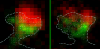
Within dendritic spines, actin is presumed to anchor receptors in the postsynaptic density and play numerous roles regulating synaptic transmission. However, the submicron dimensions of spines have hindered examination of actin dynamics within them and prevented live-cell discrimination of perisynaptic actin filaments. Using photoactivated localization microscopy, we measured movement of individual actin molecules within living spines. Velocity of single actin molecules along filaments, an index of filament polymerization rate, was highly heterogeneous within individual spines. Most strikingly, molecular velocity was elevated in discrete, well-separated foci occurring not principally at the spine tip, but in subdomains throughout the spine, including the neck. Whereas actin velocity on filaments at the synapse was substantially elevated, at the endocytic zone there was no enhanced polymerization activity. We conclude that actin subserves spatially diverse, independently regulated processes throughout spines. Perisynaptic actin forms a uniquely dynamic structure well suited for direct, active regulation of the synapse.
Commentary: A nice application of single particle tracking PALM (sptPALM), showing the flow of actin in the spines of live cultured neurons. Since 2008, the PALM in our lab has largely become a user facility, available to outside users as well as Janelians. Grad student Nick Frost in Tom Blanpied’s group at the U. of Maryland Med School visited on a number of occasions to use the PALM, with training and assistance from Hari.
A snapshot Image Mapping Spectrometer (IMS) with high sampling density is developed for hyperspectral microscopy, measuring a datacube of dimensions 285 x 285 x 60 (x, y, lambda). The spatial resolution is approximately 0.45 microm with a FOV of 100 x 100 microm(2). The measured spectrum is from 450 nm to 650 nm and is sampled by 60 spectral channels with average sampling interval approximately 3.3 nm. The channel’s spectral resolution is approximately 8nm. The spectral imaging results demonstrate the potential of the IMS for real-time cellular fluorescence imaging.