Filter
Associated Lab
- Aso Lab (1) Apply Aso Lab filter
- Cui Lab (1) Apply Cui Lab filter
- Gonen Lab (3) Apply Gonen Lab filter
- Grigorieff Lab (1) Apply Grigorieff Lab filter
- Lee (Albert) Lab (1) Apply Lee (Albert) Lab filter
- Lippincott-Schwartz Lab (1) Apply Lippincott-Schwartz Lab filter
- Pedram Lab (1) Apply Pedram Lab filter
- Rubin Lab (1) Apply Rubin Lab filter
- Sternson Lab (1) Apply Sternson Lab filter
- Tjian Lab (1) Apply Tjian Lab filter
Publication Date
- August 23, 2012 (1) Apply August 23, 2012 filter
- August 20, 2012 (2) Apply August 20, 2012 filter
- August 17, 2012 (1) Apply August 17, 2012 filter
- August 15, 2012 (1) Apply August 15, 2012 filter
- August 14, 2012 (1) Apply August 14, 2012 filter
- August 12, 2012 (1) Apply August 12, 2012 filter
- August 9, 2012 (1) Apply August 9, 2012 filter
- August 8, 2012 (1) Apply August 8, 2012 filter
- August 1, 2012 (4) Apply August 1, 2012 filter
- Remove August 2012 filter August 2012
- Remove 2012 filter 2012
Type of Publication
13 Publications
Showing 1-10 of 13 resultsAnimals approach stimuli that predict a pleasant outcome. After the paired presentation of an odour and a reward, Drosophila melanogaster can develop a conditioned approach towards that odour. Despite recent advances in understanding the neural circuits for associative memory and appetitive motivation, the cellular mechanisms for reward processing in the fly brain are unknown. Here we show that a group of dopamine neurons in the protocerebral anterior medial (PAM) cluster signals sugar reward by transient activation and inactivation of target neurons in intact behaving flies. These dopamine neurons are selectively required for the reinforcing property of, but not a reflexive response to, the sugar stimulus. In vivo calcium imaging revealed that these neurons are activated by sugar ingestion and the activation is increased on starvation. The output sites of the PAM neurons are mainly localized to the medial lobes of the mushroom bodies (MBs), where appetitive olfactory associative memory is formed. We therefore propose that the PAM cluster neurons endow a positive predictive value to the odour in the MBs. Dopamine in insects is known to mediate aversive reinforcement signals. Our results highlight the cellular specificity underlying the various roles of dopamine and the importance of spatially segregated local circuits within the MBs.
Rab proteins are important regulators of insulin-stimulated GLUT4 translocation to the plasma membrane (PM), but the precise steps in GLUT4 trafficking modulated by particular Rab proteins remain unclear. Here, we systematically investigate the involvement of Rab proteins in GLUT4 trafficking, focusing on Rab proteins directly mediating GLUT4 storage vesicle (GSV) delivery to the PM. Using dual-color total internal reflection fluorescence (TIRF) microscopy and an insulin-responsive aminopeptidase (IRAP)-pHluorin fusion assay, we demonstrated that Rab10 directly facilitated GSV translocation to and docking at the PM. Rab14 mediated GLUT4 delivery to the PM via endosomal compartments containing transferrin receptor (TfR), whereas Rab4A, Rab4B, and Rab8A recycled GLUT4 through the endosomal system. Myosin-Va associated with GSVs by interacting with Rab10, positioning peripherally recruited GSVs for ultimate fusion. Thus, multiple Rab proteins regulate the trafficking of GLUT4, with Rab10 coordinating with myosin-Va to mediate the final steps of insulin-stimulated GSV translocation to the PM.
The conserved Ndc80 complex is an essential microtubule-binding component of the kinetochore. Recent findings suggest that the Ndc80 complex influences microtubule dynamics at kinetochores in vivo. However, it was unclear if the Ndc80 complex mediates these effects directly, or by affecting other factors localized at the kinetochore. Using a reconstituted system in vitro, we show that the human Ndc80 complex directly stabilizes the tips of disassembling microtubules and promotes rescue (the transition from microtubule shortening to growth). In vivo, an N-terminal domain in the Ndc80 complex is phosphorylated by the Aurora B kinase. Mutations that mimic phosphorylation of the Ndc80 complex prevent stable kinetochore-microtubule attachment, and mutations that block phosphorylation damp kinetochore oscillations. We find that the Ndc80 complex with Aurora B phosphomimetic mutations is defective at promoting microtubule rescue, even when robustly coupled to disassembling microtubule tips. This impaired ability to affect dynamics is not simply because of weakened microtubule binding, as an N-terminally truncated complex with similar binding affinity is able to promote rescue. Taken together, these results suggest that in addition to regulating attachment stability, Aurora B controls microtubule dynamics through phosphorylation of the Ndc80 complex.
The origin of the spatial receptive fields of hippocampal place cells has not been established. A hippocampal CA1 pyramidal cell receives thousands of synaptic inputs, mostly from other spatially tuned neurons; however, how the postsynaptic neuron’s cellular properties determine the response to these inputs during behavior is unknown. We discovered that, contrary to expectations from basic models of place cells and neuronal integration, a small, spatially uniform depolarization of the spatially untuned somatic membrane potential of a silent cell leads to the sudden and reversible emergence of a spatially tuned subthreshold response and place-field spiking. Such gating of inputs by postsynaptic neuronal excitability reveals a cellular mechanism for receptive field origin and may be critical for the formation of hippocampal memory representations.
Recent behavioral studies have given rise to two contrasting models for limited working memory capacity: a "discrete-slot" model in which memory items are stored in a limited number of slots, and a "shared-resource" model in which the neural representation of items is distributed across a limited pool of resources. To elucidate the underlying neural processes, we investigated a continuous network model for working memory of an analog feature. Our model network fundamentally operates with a shared resource mechanism, and stimuli in cue arrays are encoded by a distributed neural population. On the other hand, the network dynamics and performance are also consistent with the discrete-slot model, because multiple objects are maintained by distinct localized population persistent activity patterns (bump attractors). We identified two phenomena of recurrent circuit dynamics that give rise to limited working memory capacity. As the working memory load increases, a localized persistent activity bump may either fade out (so the memory of the corresponding item is lost) or merge with another nearby bump (hence the resolution of mnemonic representation for the merged items becomes blurred). We identified specific dependences of these two phenomena on the strength and tuning of recurrent synaptic excitation, as well as network normalization: the overall population activity is invariant to set size and delay duration; therefore, a constant neural resource is shared by and dynamically allocated to the memorized items. We demonstrate that the model reproduces salient observations predicted by both discrete-slot and shared-resource models, and propose testable predictions of the merging phenomenon.
Background: It is unclear whether long-term seizure outcomes in children are similar to those in adult epilepsy surgery patients. Objective: To determine 5-year outcomes and antiepilepsy drug (AED) use in pediatric epilepsy surgery patients from a single institution. Methods: The cohort consisted of children younger than 18 years of age whose 5-year outcome data would have been available by 2010. Comparisons were made between patients with and without 5-year data (n = 338), patients with 5-year data for seizure outcome (n = 257), and seizure-free patients on and off AEDs (n = 137). Results: Five-year data were available from 76% of patients. More seizure-free patients with focal resections for hippocampal sclerosis and tumors lacked 5-year data compared with other cases. Of those with 5-year data, 53% were continuously seizure free, 18% had late seizure recurrence, 3% became seizure free after initial failure, and 25% were never seizure free. Patients were more likely to be continuously seizure free if their surgery was performed during the period 2001 to 2005 (68%) compared with surgery performed from 1996 to 2000 (61%), 1991 to 1995 (36%), and 1986 to 1990 (46%). More patients had 1 or fewer seizures per month in the late seizure recurrence (47%) compared with the not seizure-free group (20%). Four late deaths occurred in the not seizure-free group compared with 1 in the seizure-free group. Of patients who were continuously seizure free, 55% were not taking AEDs, and more cortical dysplasia patients (74%) had stopped taking AEDs compared with hemimegalencephaly patients (18%). Conclusion: In children, 5-year outcomes improved over 20 years of clinical experience. Our results are similar to those of adult epilepsy surgery patients despite mostly extratemporal and hemispheric operations for diverse developmental etiologies.
Chromosomes must be accurately partitioned to daughter cells to prevent aneuploidy, a hallmark of many tumors and birth defects. Kinetochores are the macromolecular machines that segregate chromosomes by maintaining load-bearing attachments to the dynamic tips of microtubules. Here, we present the structure of isolated budding-yeast kinetochore particles, as visualized by EM and electron tomography of negatively stained preparations. The kinetochore appears as an 126-nm particle containing a large central hub surrounded by multiple outer globular domains. In the presence of microtubules, some particles also have a ring that encircles the microtubule. Our data, showing that kinetochores bind to microtubules via multivalent attachments, lay the foundation to uncover the key mechanical and regulatory mechanisms by which kinetochores control chromosome segregation and cell division.
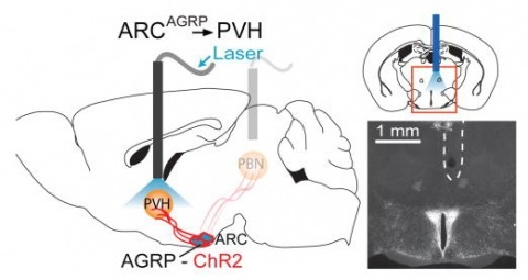
Hunger is a complex behavioural state that elicits intense food seeking and consumption. These behaviours are rapidly recapitulated by activation of starvation-sensitive AGRP neurons, which present an entry point for reverse-engineering neural circuits for hunger. Here we mapped synaptic interactions of AGRP neurons with multiple cell populations in mice and probed the contribution of these distinct circuits to feeding behaviour using optogenetic and pharmacogenetic techniques. An inhibitory circuit with paraventricular hypothalamus (PVH) neurons substantially accounted for acute AGRP neuron-evoked eating, whereas two other prominent circuits were insufficient. Within the PVH, we found that AGRP neurons target and inhibit oxytocin neurons, a small population that is selectively lost in Prader-Willi syndrome, a condition involving insatiable hunger. By developing strategies for evaluating molecularly defined circuits, we show that AGRP neuron suppression of oxytocin neurons is critical for evoked feeding. These experiments reveal a new neural circuit that regulates hunger state and pathways associated with overeating disorders.
Bacteriophage HK97 maturation involves discrete intermediate particle forms, comparable to transitional states in protein folding, before reaching its mature form. The process starts by formation of a metastable prohead, poised for exothermic expansion triggered by DNA packaging. During maturation, the capsid subunit transitions from a strained to a canonical tertiary conformation and this has been postulated to be the driving mechanism for initiating expansion via switching hexameric capsomer architecture from skewed to 6-fold symmetric. We report the subnanometer electron-cryomicroscopy reconstruction of the HK97 first expansion intermediate before any crosslink formation. This form displays 6-fold symmetric hexamers, but capsid subunit tertiary structures exhibit distortions comparable to the prohead forms. We propose that coat subunit strain release acts in synergy with the first crosslinks to drive forward maturation. Finally, we speculate that the energetic features of this transition may result from increased stability of intermediates during maturation via enhanced inter-subunit interactions.
The CA3 and CA1 pyramidal neurons are the major principal cell types of the hippocampus proper. The strongly recurrent collateral system of CA3 cells and the largely parallel-organized CA1 neurons suggest that these regions perform distinct computations. However, a comprehensive comparison between CA1 and CA3 pyramidal cells in terms of firing properties, network dynamics, and behavioral correlations is sparse in the intact animal. We performed large-scale recordings in the dorsal hippocampus of rats to quantify the similarities and differences between CA1 (n > 3,600) and CA3 (n > 2,200) pyramidal cells during sleep and exploration in multiple environments. CA1 and CA3 neurons differed significantly in firing rates, spike burst propensity, spike entrainment by the theta rhythm, and other aspects of spiking dynamics in a brain state-dependent manner. A smaller proportion of CA3 than CA1 cells displayed prominent place fields, but place fields of CA3 neurons were more compact, more stable, and carried more spatial information per spike than those of CA1 pyramidal cells. Several other features of the two cell types were specific to the testing environment. CA3 neurons showed less pronounced phase precession and a weaker position versus spike-phase relationship than CA1 cells. Our findings suggest that these distinct activity dynamics of CA1 and CA3 pyramidal cells support their distinct computational roles.