Filter
Associated Lab
Publication Date
- December 2014 (1) Apply December 2014 filter
- November 2014 (1) Apply November 2014 filter
- October 2014 (1) Apply October 2014 filter
- September 2014 (1) Apply September 2014 filter
- August 2014 (1) Apply August 2014 filter
- July 2014 (2) Apply July 2014 filter
- March 2014 (1) Apply March 2014 filter
- Remove 2014 filter 2014
Type of Publication
8 Publications
Showing 1-8 of 8 results
The origin of chordates has been debated for more than a century, with one key issue being the emergence of the notochord. In vertebrates, the notochord develops by convergence and extension of the chordamesoderm, a population of midline cells of unique molecular identity. We identify a population of mesodermal cells in a developing invertebrate, the marine annelid Platynereis dumerilii, that converges and extends toward the midline and expresses a notochord-specific combination of genes. These cells differentiate into a longitudinal muscle, the axochord, that is positioned between central nervous system and axial blood vessel and secretes a strong collagenous extracellular matrix. Ancestral state reconstruction suggests that contractile mesodermal midline cells existed in bilaterian ancestors. We propose that these cells, via vacuolization and stiffening, gave rise to the chordate notochord.
The comprehensive reconstruction of cell lineages in complex multicellular organisms is a central goal of developmental biology. We present an open-source computational framework for the segmentation and tracking of cell nuclei with high accuracy and speed. We demonstrate its (i) generality by reconstructing cell lineages in four-dimensional, terabyte-sized image data sets of fruit fly, zebrafish and mouse embryos acquired with three types of fluorescence microscopes, (ii) scalability by analyzing advanced stages of development with up to 20,000 cells per time point at 26,000 cells min(-1) on a single computer workstation and (iii) ease of use by adjusting only two parameters across all data sets and providing visualization and editing tools for efficient data curation. Our approach achieves on average 97.0% linkage accuracy across all species and imaging modalities. Using our system, we performed the first cell lineage reconstruction of early Drosophila melanogaster nervous system development, revealing neuroblast dynamics throughout an entire embryo.
The fruit fly is an excellent model system for investigating the sequence of epithelial tissue invaginations constituting the process of gastrulation. By combining recent advancements in light sheet fluorescence microscopy (LSFM) and image processing, the three-dimensional fly embryo morphology and relevant gene expression patterns can be accurately recorded throughout the entire process of embryogenesis. LSFM provides exceptionally high imaging speed, high signal-to-noise ratio, low level of photoinduced damage, and good optical penetration depth. This powerful combination of capabilities makes LSFM particularly suitable for live imaging of the fly embryo.The resulting high-information-content image data are subsequently processed to obtain the outlines of cells and cell nuclei, as well as the geometry of the whole embryo tissue by image segmentation. Furthermore, morphodynamics information is extracted by computationally tracking objects in the image. Towards that goal we describe the successful implementation of a fast fitting strategy of Gaussian mixture models.The data obtained by image processing is well-suited for hypothesis testing of the detailed biomechanics of the gastrulating embryo. Typically this involves constructing computational mechanics models that consist of an objective function providing an estimate of strain energy for a given morphological configuration of the tissue, and a numerical minimization mechanism of this energy, achieved by varying morphological parameters.In this chapter, we provide an overview of in vivo imaging of fruit fly embryos using LSFM, computational tools suitable for processing the resulting images, and examples of computational biomechanical simulations of fly embryo gastrulation.
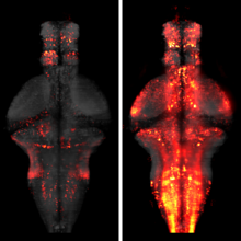
The processing of sensory input and the generation of behavior involves large networks of neurons, which necessitates new technology for recording from many neurons in behaving animals. In the larval zebrafish, light-sheet microscopy can be used to record the activity of almost all neurons in the brain simultaneously at single-cell resolution. Existing implementations, however, cannot be combined with visually driven behavior because the light sheet scans over the eye, interfering with presentation of controlled visual stimuli. Here we describe a system that overcomes the confounding eye stimulation through the use of two light sheets and combines whole-brain light-sheet imaging with virtual reality for fictively behaving larval zebrafish.
Developments in electrical and optical recording technology are scaling up the size of neuronal populations that can be monitored simultaneously. Light-sheet imaging is rapidly gaining traction as a method for optically interrogating activity in large networks and presents both opportunities and challenges for understanding circuit function.
This protocol describes how to observe gastrulation in living mouse embryos by using light-sheet microscopy and computational tools to analyze the resulting image data at the single-cell level. We describe a series of techniques needed to image the embryos under physiological conditions, including how to hold mouse embryos without agarose embedding, how to transfer embryos without air exposure and how to construct environmental chambers for live imaging by digital scanned light-sheet microscopy (DSLM). Computational tools include manual and semiautomatic tracking programs that are developed for analyzing the large 4D data sets acquired with this system. Note that this protocol does not include details of how to build the light-sheet microscope itself. Time-lapse imaging ends within 12 h, with subsequent tracking analysis requiring 3-6 d. Other than some mouse-handling skills, this protocol requires no advanced skills or knowledge. Light-sheet microscopes are becoming more widely available, and thus the techniques outlined in this paper should be helpful for investigating mouse embryogenesis.
The molecular and cellular architecture of the organs in a whole mouse is revealed through optical clearing.

Inflammatory cells acquire a polarized phenotype to migrate towards sites of infection or injury. A conserved polarity complex comprising PAR-3, PAR-6 and atypical protein kinase C (aPKC) relays extracellular polarizing cues to control cytoskeletal and signaling networks affecting morphological and functional polarization. However, there is no evidence that myeloid cells use PAR signaling to migrate vectorially in three-dimensional (3D) environments in vivo. Using genetically encoded bioprobes and high-resolution live imaging, we reveal the existence of F-actin oscillations in the trailing edge and constant repositioning of the microtubule organizing center (MTOC) to direct leukocyte migration in wounded medaka fish larvae (Oryzias latipes). Genetic manipulation in live myeloid cells demonstrates that the catalytic activity of aPKC and the regulated interaction with PAR-3 and PAR-6 are required for consistent F-actin oscillations, MTOC perinuclear mobility, aPKC repositioning and wound-directed migration upstream of Rho kinase (also known as ROCK or ROK) activation. We propose that the PAR complex coordinately controls cytoskeletal changes affecting both the generation of traction force and the directionality of leukocyte migration to sites of injury.