Filter
Associated Lab
- Ahrens Lab (4) Apply Ahrens Lab filter
- Betzig Lab (2) Apply Betzig Lab filter
- Branson Lab (1) Apply Branson Lab filter
- Darshan Lab (3) Apply Darshan Lab filter
- Druckmann Lab (5) Apply Druckmann Lab filter
- Dudman Lab (3) Apply Dudman Lab filter
- Fetter Lab (1) Apply Fetter Lab filter
- Freeman Lab (3) Apply Freeman Lab filter
- Harris Lab (6) Apply Harris Lab filter
- Hermundstad Lab (1) Apply Hermundstad Lab filter
- Jayaraman Lab (9) Apply Jayaraman Lab filter
- Ji Lab (2) Apply Ji Lab filter
- Karpova Lab (1) Apply Karpova Lab filter
- Lavis Lab (4) Apply Lavis Lab filter
- Lee (Albert) Lab (3) Apply Lee (Albert) Lab filter
- Leonardo Lab (2) Apply Leonardo Lab filter
- Liu (Zhe) Lab (1) Apply Liu (Zhe) Lab filter
- Looger Lab (20) Apply Looger Lab filter
- Magee Lab (1) Apply Magee Lab filter
- Pachitariu Lab (2) Apply Pachitariu Lab filter
- Podgorski Lab (2) Apply Podgorski Lab filter
- Romani Lab (5) Apply Romani Lab filter
- Rubin Lab (3) Apply Rubin Lab filter
- Saalfeld Lab (2) Apply Saalfeld Lab filter
- Schreiter Lab (13) Apply Schreiter Lab filter
- Spruston Lab (3) Apply Spruston Lab filter
- Sternson Lab (4) Apply Sternson Lab filter
- Remove Svoboda Lab filter Svoboda Lab
- Tillberg Lab (3) Apply Tillberg Lab filter
- Turner Lab (3) Apply Turner Lab filter
Associated Project Team
Associated Support Team
- Anatomy and Histology (4) Apply Anatomy and Histology filter
- Gene Targeting and Transgenics (3) Apply Gene Targeting and Transgenics filter
- Integrative Imaging (2) Apply Integrative Imaging filter
- Janelia Experimental Technology (4) Apply Janelia Experimental Technology filter
- Molecular Genomics (2) Apply Molecular Genomics filter
- Primary & iPS Cell Culture (2) Apply Primary & iPS Cell Culture filter
- Project Technical Resources (1) Apply Project Technical Resources filter
- Quantitative Genomics (1) Apply Quantitative Genomics filter
- Scientific Computing Software (2) Apply Scientific Computing Software filter
- Viral Tools (2) Apply Viral Tools filter
Publication Date
- 2023 (5) Apply 2023 filter
- 2022 (6) Apply 2022 filter
- 2021 (7) Apply 2021 filter
- 2020 (5) Apply 2020 filter
- 2019 (14) Apply 2019 filter
- 2018 (11) Apply 2018 filter
- 2017 (9) Apply 2017 filter
- 2016 (8) Apply 2016 filter
- 2015 (9) Apply 2015 filter
- 2014 (7) Apply 2014 filter
- 2013 (10) Apply 2013 filter
- 2012 (9) Apply 2012 filter
- 2011 (7) Apply 2011 filter
- 2010 (7) Apply 2010 filter
- 2009 (8) Apply 2009 filter
- 2008 (6) Apply 2008 filter
- 2007 (3) Apply 2007 filter
131 Janelia Publications
Showing 71-80 of 131 resultsMany forms of human and animal behavior involve head movements. A new study reveals the neural code for three-dimensional head movements in the midbrain of freely moving mice.
The mechanisms linking sensation and action during learning are poorly understood. Layer 2/3 neurons in the motor cortex might participate in sensorimotor integration and learning; they receive input from sensory cortex and excite deep layer neurons, which control movement. Here we imaged activity in the same set of layer 2/3 neurons in the motor cortex over weeks, while mice learned to detect objects with their whiskers and report detection with licking. Spatially intermingled neurons represented sensory (touch) and motor behaviours (whisker movements and licking). With learning, the population-level representation of task-related licking strengthened. In trained mice, population-level representations were redundant and stable, despite dynamism of single-neuron representations. The activity of a subpopulation of neurons was consistent with touch driving licking behaviour. Our results suggest that ensembles of motor cortex neurons couple sensory input to multiple, related motor programs during learning.
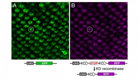
Site-specific recombinases have been used for two decades to manipulate the structure of animal genomes in highly predictable ways and have become major research tools. However, the small number of recombinases demonstrated to have distinct specificities, low toxicity, and sufficient activity to drive reactions to completion in animals has been a limitation. In this report we show that four recombinases derived from yeast-KD, B2, B3, and R-are highly active and nontoxic in Drosophila and that KD, B2, B3, and the widely used FLP recombinase have distinct target specificities. We also show that the KD and B3 recombinases are active in mice.
The distinct electrical properties of axonal and dendritic membranes are largely a result of specific transport of vesicle-bound membrane proteins to each compartment. How this specificity arises is unclear because kinesin motors that transport vesicles cannot autonomously distinguish dendritically projecting microtubules from those projecting axonally. We hypothesized that interaction with a second motor might enable vesicles containing dendritic proteins to preferentially associate with dendritically projecting microtubules and avoid those that project to the axon. Here we show that in rat cortical neurons, localization of several distinct transmembrane proteins to dendrites is dependent on specific myosin motors and an intact actin network. Moreover, fusion with a myosin-binding domain from Melanophilin targeted Channelrhodopsin-2 specifically to the somatodendritic compartment of neurons in mice in vivo. Together, our results suggest that dendritic transmembrane proteins direct the vesicles in which they are transported to avoid the axonal compartment through interaction with myosin motors.
During many natural behaviors the relevant sensory stimuli and motor outputs are difficult to quantify. Furthermore, the high dimensionality of the space of possible stimuli and movements compounds the problem of experimental control. Head fixation facilitates stimulus control and movement tracking, and can be combined with techniques for recording and manipulating neural activity. However, head-fixed mouse behaviors are typically trained through extensive instrumental conditioning. Here we present a whisker-based, tactile virtual reality system for head-fixed mice running on a spherical treadmill. Head-fixed mice displayed natural movements, including running and rhythmic whisking at 16 Hz. Whisking was centered on a set point that changed in concert with running so that more protracted whisking was correlated with faster running. During turning, whiskers moved in an asymmetric manner, with more retracted whisker positions in the turn direction and protracted whisker movements on the other side. Under some conditions, whisker movements were phase-coupled to strides. We simulated a virtual reality tactile corridor, consisting of two moveable walls controlled in a closed-loop by running speed and direction. Mice used their whiskers to track the walls of the winding corridor without training. Whisker curvature changes, which cause forces in the sensory follicles at the base of the whiskers, were tightly coupled to distance from the walls. Our behavioral system allows for precise control of sensorimotor variables during natural tactile navigation.
Classical studies have related the spiking of selected neocortical neurons to behavior, but little is known about activity sampled from the entire neural population. We recorded from neurons selected independent of spiking, using cell-attached recordings and two-photon calcium imaging, in the barrel cortex of mice performing an object localization task. Spike rates varied across neurons, from silence to >60 Hz. Responses were diverse, with some neurons showing large increases in spike rate when whiskers contacted the object. Nearly half the neurons discriminated object location; a small fraction of neurons discriminated perfectly. More active neurons were more discriminative. Layer (L) 4 and L5 contained the highest fractions of discriminating neurons (\~{}63% and 79%, respectively), but a few L2/3 neurons were also highly discriminating. Approximately 13,000 spikes per activated barrel column were available to mice for decision making. Coding of object location in the barrel cortex is therefore highly redundant.
The brain plans and executes volitional movements. The underlying patterns of neural population activity have been explored in the context of movements of the eyes, limbs, tongue, and head in nonhuman primates and rodents. How do networks of neurons produce the slow neural dynamics that prepare specific movements and the fast dynamics that ultimately initiate these movements? Recent work exploits rapid and calibrated perturbations of neural activity to test specific dynamical systems models that are capable of producing the observed neural activity. These joint experimental and computational studies show that cortical dynamics during motor planning reflect fixed points of neural activity (attractors). Subcortical control signals reshape and move attractors over multiple timescales, causing commitment to specific actions and rapid transitions to movement execution. Experiments in rodents are beginning to reveal how these algorithms are implemented at the level of brain-wide neural circuits. Expected final online publication date for the , Volume 45 is July 2022. Please see http://www.annualreviews.org/page/journal/pubdates for revised estimates.
The brain plans and executes volitional movements. The underlying patterns of neural population activity have been explored in the context of movements of the eyes, limbs, tongue, and head in nonhuman primates and rodents. How do networks of neurons produce the slow neural dynamics that prepare specific movements and the fast dynamics that ultimately initiate these movements? Recent work exploits rapid and calibrated perturbations of neural activity to test specific dynamical systems models that are capable of producing the observed neural activity. These joint experimental and computational studies show that cortical dynamics during motor planning reflect fixed points of neural activity (attractors). Subcortical control signals reshape and move attractors over multiple timescales, causing commitment to specific actions and rapid transitions to movement execution. Experiments in rodents are beginning to reveal how these algorithms are implemented at the level of brain-wide neural circuits.
Active sensation requires the convergence of external stimuli with representations of body movements. We used mouse behavior, electrophysiology and optogenetics to dissect the temporal interactions among whisker movement, neural activity and sensation of touch. We photostimulated layer 4 activity in single barrels in a closed loop with whisking. Mimicking touch-related neural activity caused illusory perception of an object at a particular location, but scrambling the timing of the spikes over one whisking cycle (tens of milliseconds) did not abolish the illusion, indicating that knowledge of instantaneous whisker position is unnecessary for discriminating object locations. The illusions were induced only during bouts of directed whisking, when mice expected touch, and in the relevant barrel. Reducing activity biased behavior, consistent with a spike count code for object detection at a particular location. Our results show that mice integrate coding of touch with movement over timescales of a whisking bout to produce perception of active touch.
Animals seek out relevant information by moving through a dynamic world, but sensory systems are usually studied under highly constrained and passive conditions that may not probe important dimensions of the neural code. Here, we explored neural coding in the barrel cortex of head-fixed mice that tracked walls with their whiskers in tactile virtual reality. Optogenetic manipulations revealed that barrel cortex plays a role in wall-tracking. Closed-loop optogenetic control of layer 4 neurons can substitute for whisker-object contact to guide behavior resembling wall tracking. We measured neural activity using two-photon calcium imaging and extracellular recordings. Neurons were tuned to the distance between the animal snout and the contralateral wall, with monotonic, unimodal, and multimodal tuning curves. This rich representation of object location in the barrel cortex could not be predicted based on simple stimulus-response relationships involving individual whiskers and likely emerges within cortical circuits.