Filter
Associated Lab
- Ahrens Lab (7) Apply Ahrens Lab filter
- Druckmann Lab (2) Apply Druckmann Lab filter
- Dudman Lab (1) Apply Dudman Lab filter
- Freeman Lab (2) Apply Freeman Lab filter
- Harris Lab (7) Apply Harris Lab filter
- Hermundstad Lab (1) Apply Hermundstad Lab filter
- Hess Lab (2) Apply Hess Lab filter
- Jayaraman Lab (10) Apply Jayaraman Lab filter
- Karpova Lab (2) Apply Karpova Lab filter
- Keller Lab (5) Apply Keller Lab filter
- Lavis Lab (8) Apply Lavis Lab filter
- Leonardo Lab (4) Apply Leonardo Lab filter
- Remove Looger Lab filter Looger Lab
- Podgorski Lab (6) Apply Podgorski Lab filter
- Rubin Lab (2) Apply Rubin Lab filter
- Schreiter Lab (24) Apply Schreiter Lab filter
- Simpson Lab (1) Apply Simpson Lab filter
- Spruston Lab (1) Apply Spruston Lab filter
- Sternson Lab (2) Apply Sternson Lab filter
- Svoboda Lab (20) Apply Svoboda Lab filter
- Tervo Lab (1) Apply Tervo Lab filter
- Tillberg Lab (1) Apply Tillberg Lab filter
- Turner Lab (1) Apply Turner Lab filter
- Zlatic Lab (1) Apply Zlatic Lab filter
Associated Project Team
Associated Support Team
- Anatomy and Histology (2) Apply Anatomy and Histology filter
- Cryo-Electron Microscopy (1) Apply Cryo-Electron Microscopy filter
- Electron Microscopy (1) Apply Electron Microscopy filter
- Fly Facility (1) Apply Fly Facility filter
- Janelia Experimental Technology (2) Apply Janelia Experimental Technology filter
- Molecular Genomics (1) Apply Molecular Genomics filter
- Primary & iPS Cell Culture (2) Apply Primary & iPS Cell Culture filter
- Quantitative Genomics (2) Apply Quantitative Genomics filter
- Scientific Computing Software (1) Apply Scientific Computing Software filter
- Viral Tools (1) Apply Viral Tools filter
- Vivarium (1) Apply Vivarium filter
Publication Date
- 2024 (1) Apply 2024 filter
- 2023 (5) Apply 2023 filter
- 2022 (7) Apply 2022 filter
- 2021 (11) Apply 2021 filter
- 2020 (7) Apply 2020 filter
- 2019 (15) Apply 2019 filter
- 2018 (8) Apply 2018 filter
- 2017 (6) Apply 2017 filter
- 2016 (10) Apply 2016 filter
- 2015 (9) Apply 2015 filter
- 2014 (11) Apply 2014 filter
- 2013 (10) Apply 2013 filter
- 2012 (13) Apply 2012 filter
- 2011 (7) Apply 2011 filter
- 2010 (6) Apply 2010 filter
- 2009 (7) Apply 2009 filter
- 2008 (3) Apply 2008 filter
136 Janelia Publications
Showing 81-90 of 136 resultsA fundamental question in sensory neuroscience is how parallel processing is implemented at the level of molecular and circuit mechanisms. In the retina, it has been proposed that distinct OFF cone bipolar cell types generate fast/transient and slow/sustained pathways by the differential expression of AMPA- and kainate-type glutamate receptors, respectively. However, the functional significance of these receptors in the intact circuit during light stimulation remains unclear. Here, we measured glutamate release from mouse bipolar cells by two-photon imaging of a glutamate sensor (iGluSnFR) expressed on postsynaptic amacrine and ganglion cell dendrites. In both transient and sustained OFF layers, cone-driven glutamate release from bipolar cells was blocked by antagonists to kainate receptors but not AMPA receptors. Electrophysiological recordings from bipolar and ganglion cells confirmed the essential role of kainate receptors for signaling in both transient and sustained OFF pathways. Kainate receptors mediated responses to contrast modulation up to 20 Hz. Light-evoked responses in all mouse OFF bipolar pathways depend on kainate, not AMPA, receptors.
The brain can become transiently disconnected from the environment while maintaining vivid, internally generated experiences. This so-called 'dissociated state' can occur in pathological conditions and under the influence of psychedelics or the anesthetic ketamine (KET). The cellular and circuit mechanisms producing the dissociative state remain poorly understood. We show in mice that KET causes spontaneously active neurons to become suppressed while previously silent neurons become spontaneously activated. This switch occurs in all cortical layers and different cortical regions, is induced by both systemic and cortical application of KET and is mediated by suppression of parvalbumin and somatostatin interneuron activity and inhibition of NMDA receptors and HCN channels. Combined, our results reveal two largely non-overlapping cortical neuronal populations-one engaged in wakefulness, the other contributing to the KET-induced brain state-and may lay the foundation for understanding how the brain might become disconnected from the surrounding environment while maintaining internal subjective experiences.
Point-scanning two-photon microscopy enables high-resolution imaging within scattering specimens such as the mammalian brain, but sequential acquisition of voxels fundamentally limits imaging speed. We developed a two-photon imaging technique that scans lines of excitation across a focal plane at multiple angles and uses prior information to recover high-resolution images at over 1.4 billion voxels per second. Using a structural image as a prior for recording neural activity, we imaged visually-evoked and spontaneous glutamate release across hundreds of dendritic spines in mice at depths over 250 microns and frame-rates over 1 kHz. Dendritic glutamate transients in anaesthetized mice are synchronized within spatially-contiguous domains spanning tens of microns at frequencies ranging from 1-100 Hz. We demonstrate high-speed recording of acetylcholine and calcium sensors, 3D single-particle tracking, and imaging in densely-labeled cortex. Our method surpasses limits on the speed of raster-scanned imaging imposed by fluorescence lifetime.
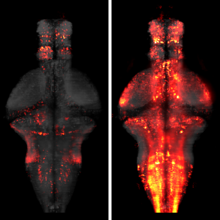
The identification of active neurons and circuits in vivo is a fundamental challenge in understanding the neural basis of behavior. Genetically encoded calcium (Ca(2+)) indicators (GECIs) enable quantitative monitoring of cellular-resolution activity during behavior. However, such indicators require online monitoring within a limited field of view. Alternatively, post hoc staining of immediate early genes (IEGs) indicates highly active cells within the entire brain, albeit with poor temporal resolution. We designed a fluorescent sensor, CaMPARI, that combines the genetic targetability and quantitative link to neural activity of GECIs with the permanent, large-scale labeling of IEGs, allowing a temporally precise "activity snapshot" of a large tissue volume. CaMPARI undergoes efficient and irreversible green-to-red conversion only when elevated intracellular Ca(2+) and experimenter-controlled illumination coincide. We demonstrate the utility of CaMPARI in freely moving larvae of zebrafish and flies, and in head-fixed mice and adult flies.
Photoreceptors for visual perception, phototaxis or light avoidance are typically clustered in eyes or related structures such as the Bolwig organ of Drosophila larvae. Unexpectedly, we found that the class IV dendritic arborization neurons of Drosophila melanogaster larvae respond to ultraviolet, violet and blue light, and are major mediators of light avoidance, particularly at high intensities. These class IV dendritic arborization neurons, which are present in every body segment, have dendrites tiling the larval body wall nearly completely without redundancy. Dendritic illumination activates class IV dendritic arborization neurons. These novel photoreceptors use phototransduction machinery distinct from other photoreceptors in Drosophila and enable larvae to sense light exposure over their entire bodies and move out of danger.
The processing of sensory input and the generation of behavior involves large networks of neurons, which necessitates new technology for recording from many neurons in behaving animals. In the larval zebrafish, light-sheet microscopy can be used to record the activity of almost all neurons in the brain simultaneously at single-cell resolution. Existing implementations, however, cannot be combined with visually driven behavior because the light sheet scans over the eye, interfering with presentation of controlled visual stimuli. Here we describe a system that overcomes the confounding eye stimulation through the use of two light sheets and combines whole-brain light-sheet imaging with virtual reality for fictively behaving larval zebrafish.
OBJECTIVE: A recent genome-wide association study (GWAS) reported a significant genetic association between rs34330 of cyclin-dependent kinase inhibitor 1B (CDKN1B) and risk of systemic lupus erythematosus (SLE) in Han Chinese. This study aims to validate the reported association and elucidate the biochemical mechanisms underlying the variant's effect. METHODS: We performed allelic association with SLE followed by meta-analysis across 11 independent cohorts (n=28,872). We applied in silico bioinformatics and experimental validation in SLE-relevant cell lines to determine the functional consequences of rs34330. RESULTS: We replicated genetic association between SLE and rs34330 (P =5.29x10 , OR (95% CI)=0.84 (0.81-0.87)). Follow-up bioinformatics and eQTL analysis suggest that rs34330 is located in active chromatin and potentially regulates several target genes. Using luciferase and ChIP-qPCR, we demonstrated substantial allele-specific promoter and enhancer activity, and allele-specific binding of three histone marks (H3K27ac, H3K4me3, H3K4me1), RNA pol II, CTCF, and a critical immune transcription factor (IRF-1). Chromosome conformation capture (3C) detected long-range chromatin interactions between rs34330 and the promoters of neighboring genes APOLD1 and DDX47, and effects on CDKN1B and the other target genes were directly validated by CRISPR-based genome editing. Finally, CRISPR-dCas9-based epigenetic activation/silencing confirmed these results. Gene-edited cell lines also showed higher levels of proliferation and apoptosis. CONCLUSION: Collectively, these findings suggest a mechanism whereby the rs34330 risk allele (C) influences the presence of histone marks, RNA pol II, and the IRF-1 transcription factor to regulate expression of several target genes linked to proliferation and apoptosis, which potentially underlie the association of rs34330 with SLE.
Hundreds of millions of structured proteins sustain life through chemical interactions and catalytic reactions1. Though dynamic, these proteins are assumed to be built upon fixed scaffolds of secondary structure, α-helices and β-sheets. Experimentally determined structures of over >58,000 non-redundant proteins support this assumption, though it has recently been challenged by ∼100 fold-switching proteins2. These “metamorphic3” proteins, though ostensibly rare, raise the question of how many uncharacterized proteins have shapeshifting–rather than fixed–secondary structures. To address this question, we developed a comparative sequence-based approach that predicts fold-switching proteins from differences in secondary structure propensity. We applied this approach to the universally conserved NusG transcription factor family of ∼15,000 proteins, one of which has a 50-residue regulatory subunit experimentally shown to switch between α-helical and β-sheet folds4. Our approach predicted that 25% of the sequences in this family undergo similar α-helix ⇌ β-sheet transitions, a frequency two orders of magnitude larger than previously observed. Our predictions evade state-of-the-art computational methods but were confirmed experimentally by circular dichroism and nuclear magnetic resonance spectroscopy for all 10 assiduously chosen dissimilar variants. These results suggest that fold switching is a pervasive mechanism of transcriptional regulation in all kingdoms of life and imply that numerous uncharacterized proteins may also switch folds.
The protein folding paradigm asserts that the three-dimensional structure of a protein is determined by its amino acid sequence. Here we show that a substantial population of proteins from the NusG superfamily of transcription factors do not adhere to this paradigm. Previous work demonstrated that one member of this superfamily has a regulatory domain that completely switches between α-helical and β-sheet folds, but the pervasiveness of this fold-switching mechanism is uncertain. To address this question, we developed a sequence-based predictor, which revealed that thousands of proteins from this superfamily switch folds. Circular dichroism and nuclear magnetic resonance spectroscopies of 10 sequence-diverse variants confirmed our predictions. By contrast, state-of-the-art methods based on the protein folding paradigm assume that related sequences adopt the same fold and thus predicted that the regulatory domains of all variants adopt only the β-sheet fold. Removal of this bias revealed that residue-residue contacts from both α-helical and β-sheet folds are conserved in a large subpopulation of fold-switching domains, poising them to assume disparate conformations. Our results suggest that fold switching is a pervasive mechanism of transcriptional regulation in all kingdoms of life and indicate that expanding the protein folding paradigm may reveal the involvement of fold-switching proteins in diverse biological processes.