Filter
Associated Lab
- Ahrens Lab (5) Apply Ahrens Lab filter
- Branson Lab (3) Apply Branson Lab filter
- Card Lab (1) Apply Card Lab filter
- Cardona Lab (1) Apply Cardona Lab filter
- Druckmann Lab (1) Apply Druckmann Lab filter
- Freeman Lab (4) Apply Freeman Lab filter
- Funke Lab (2) Apply Funke Lab filter
- Harris Lab (1) Apply Harris Lab filter
- Ji Lab (1) Apply Ji Lab filter
- Remove Keller Lab filter Keller Lab
- Lavis Lab (2) Apply Lavis Lab filter
- Liu (Zhe) Lab (1) Apply Liu (Zhe) Lab filter
- Looger Lab (5) Apply Looger Lab filter
- Pavlopoulos Lab (1) Apply Pavlopoulos Lab filter
- Tillberg Lab (1) Apply Tillberg Lab filter
- Turaga Lab (1) Apply Turaga Lab filter
Associated Project Team
Associated Support Team
Publication Date
- 2024 (1) Apply 2024 filter
- 2023 (1) Apply 2023 filter
- 2022 (1) Apply 2022 filter
- 2021 (2) Apply 2021 filter
- 2020 (5) Apply 2020 filter
- 2019 (6) Apply 2019 filter
- 2018 (6) Apply 2018 filter
- 2017 (2) Apply 2017 filter
- 2016 (6) Apply 2016 filter
- 2015 (7) Apply 2015 filter
- 2014 (7) Apply 2014 filter
- 2013 (9) Apply 2013 filter
- 2012 (3) Apply 2012 filter
- 2011 (2) Apply 2011 filter
- 2010 (2) Apply 2010 filter
60 Janelia Publications
Showing 31-40 of 60 results
The fruit fly is an excellent model system for investigating the sequence of epithelial tissue invaginations constituting the process of gastrulation. By combining recent advancements in light sheet fluorescence microscopy (LSFM) and image processing, the three-dimensional fly embryo morphology and relevant gene expression patterns can be accurately recorded throughout the entire process of embryogenesis. LSFM provides exceptionally high imaging speed, high signal-to-noise ratio, low level of photoinduced damage, and good optical penetration depth. This powerful combination of capabilities makes LSFM particularly suitable for live imaging of the fly embryo.The resulting high-information-content image data are subsequently processed to obtain the outlines of cells and cell nuclei, as well as the geometry of the whole embryo tissue by image segmentation. Furthermore, morphodynamics information is extracted by computationally tracking objects in the image. Towards that goal we describe the successful implementation of a fast fitting strategy of Gaussian mixture models.The data obtained by image processing is well-suited for hypothesis testing of the detailed biomechanics of the gastrulating embryo. Typically this involves constructing computational mechanics models that consist of an objective function providing an estimate of strain energy for a given morphological configuration of the tissue, and a numerical minimization mechanism of this energy, achieved by varying morphological parameters.In this chapter, we provide an overview of in vivo imaging of fruit fly embryos using LSFM, computational tools suitable for processing the resulting images, and examples of computational biomechanical simulations of fly embryo gastrulation.
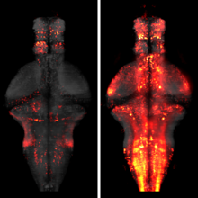
The processing of sensory input and the generation of behavior involves large networks of neurons, which necessitates new technology for recording from many neurons in behaving animals. In the larval zebrafish, light-sheet microscopy can be used to record the activity of almost all neurons in the brain simultaneously at single-cell resolution. Existing implementations, however, cannot be combined with visually driven behavior because the light sheet scans over the eye, interfering with presentation of controlled visual stimuli. Here we describe a system that overcomes the confounding eye stimulation through the use of two light sheets and combines whole-brain light-sheet imaging with virtual reality for fictively behaving larval zebrafish.
Developments in electrical and optical recording technology are scaling up the size of neuronal populations that can be monitored simultaneously. Light-sheet imaging is rapidly gaining traction as a method for optically interrogating activity in large networks and presents both opportunities and challenges for understanding circuit function.

The ability to visualize and quantitatively measure dynamic biological processes in vivo and at high spatiotemporal resolution is of fundamental importance to experimental investigations in developmental biology. Light-sheet microscopy is particularly well suited to providing such data, since it offers exceptionally high imaging speed and good spatial resolution while minimizing light-induced damage to the specimen. We review core principles and recent advances in light-sheet microscopy, with a focus on concepts and implementations relevant for applications in developmental biology. We discuss how light-sheet microcopy has helped advance our understanding of developmental processes from single-molecule to whole-organism studies, assess the potential for synergies with other state-of-the-art technologies, and introduce methods for computational image and data analysis. Finally, we explore the future trajectory of light-sheet microscopy, discuss key efforts to disseminate new light-sheet technology, and identify exciting opportunities for further advances.

In vivo imaging applications typically require carefully balancing conflicting parameters. Often it is necessary to achieve high imaging speed, low photo-bleaching, and photo-toxicity, good three-dimensional resolution, high signal-to-noise ratio, and excellent physical coverage at the same time. Light-sheet microscopy provides good performance in all of these categories, and is thus emerging as a particularly powerful live imaging method for the life sciences. We see an outstanding potential for applying light-sheet microscopy to the study of development and function of the early nervous system in vertebrates and higher invertebrates. Here, we review state-of-the-art approaches to live imaging of early development, and show how the unique capabilities of light-sheet microscopy can further advance our understanding of the development and function of the nervous system. We discuss key considerations in the design of light-sheet microscopy experiments, including sample preparation and fluorescent marker strategies, and provide an outlook for future directions in the field.
The molecular and cellular architecture of the organs in a whole mouse is revealed through optical clearing.
The thirteen nuclear cleavages that give rise to the Drosophila blastoderm are some of the fastest known cell cycles. Surprisingly, the fertilized egg is provided with at most one-third of the dNTPs needed to complete the thirteen rounds of DNA replication. The rest must be synthesized by the embryo, concurrent with cleavage divisions. What is the reason for the limited supply of DNA building blocks? We propose that frugal control of dNTP synthesis contributes to the well-characterized deceleration of the cleavage cycles and is needed for robust accumulation of zygotic gene products. In support of this model, we demonstrate that when the levels of dNTPs are abnormally high, nuclear cleavages fail to sufficiently decelerate, the levels of zygotic transcription are dramatically reduced, and the embryo catastrophically fails early in gastrulation. Our work reveals a direct connection between metabolism, the cell cycle, and zygotic transcription.

During development, coordinated cell behaviors orchestrate tissue and organ morphogenesis. Detailed descriptions of cell lineages and behaviors provide a powerful framework to elucidate the mechanisms of morphogenesis. To study the cellular basis of limb development, we imaged transgenic fluorescently-labeled embryos from the crustacean Parhyale hawaiensis with multi-view light-sheet microscopy at high spatiotemporal resolution over several days of embryogenesis. The cell lineage of outgrowing thoracic limbs was reconstructed at single-cell resolution with new software called Massive Multi-view Tracker (MaMuT). In silico clonal analyses suggested that the early limb primordium becomes subdivided into anterior-posterior and dorsal-ventral compartments whose boundaries intersect at the distal tip of the growing limb. Limb-bud formation is associated with spatial modulation of cell proliferation, while limb elongation is also driven by preferential orientation of cell divisions along the proximal-distal growth axis. Cellular reconstructions were predictive of the expression patterns of limb development genes including the BMP morphogen Decapentaplegic.
Visualization of cellular and molecular processes is an indispensable tool for cell biologists, and innovations in microscopy methods unfailingly lead to new biological discoveries. Today, light microscopy (LM) provides ever-higher spatial and temporal resolution and visualization of biological process over enormous ranges. Electron microscopy (EM) is moving into the atomic resolution regime and allowing cellular analyses that are more physiological and sophisticated in scope. Importantly, much is being gained by combining multiple approaches, (e.g., LM and EM) to take advantage of their complementary strengths. The advent of high-throughput microscopies has led to a common need for sophisticated computational methods to quantitatively analyze huge amounts of data and translate images into new biological insights.

An important question in early neural development is the origin of stochastic nuclear movement between apical and basal surfaces of neuroepithelia during interkinetic nuclear migration. Tracking of nuclear subpopulations has shown evidence of diffusion - mean squared displacements growing linearly in time - and suggested crowding from cell division at the apical surface drives basalward motion. Yet, this hypothesis has not yet been tested, and the forces involved not quantified. We employ long-term, rapid light-sheet and two-photon imaging of early zebrafish retinogenesis to track entire populations of nuclei within the tissue. The time-varying concentration profiles show clear evidence of crowding as nuclei reach close-packing and are quantitatively described by a nonlinear diffusion model. Considerations of nuclear motion constrained inside the enveloping cell membrane show that concentration-dependent stochastic forces inside cells, compatible in magnitude to those found in cytoskeletal transport, can explain the observed magnitude of the diffusion constant.