Filter
Associated Lab
- Ahrens Lab (5) Apply Ahrens Lab filter
- Beyene Lab (1) Apply Beyene Lab filter
- Branson Lab (3) Apply Branson Lab filter
- Card Lab (1) Apply Card Lab filter
- Cardona Lab (1) Apply Cardona Lab filter
- Druckmann Lab (1) Apply Druckmann Lab filter
- Freeman Lab (4) Apply Freeman Lab filter
- Funke Lab (2) Apply Funke Lab filter
- Harris Lab (1) Apply Harris Lab filter
- Ji Lab (1) Apply Ji Lab filter
- Keller Lab (76) Apply Keller Lab filter
- Lavis Lab (3) Apply Lavis Lab filter
- Liu (Zhe) Lab (1) Apply Liu (Zhe) Lab filter
- Looger Lab (5) Apply Looger Lab filter
- Pavlopoulos Lab (1) Apply Pavlopoulos Lab filter
- Schreiter Lab (1) Apply Schreiter Lab filter
- Stringer Lab (1) Apply Stringer Lab filter
- Tillberg Lab (1) Apply Tillberg Lab filter
- Turaga Lab (1) Apply Turaga Lab filter
- Turner Lab (1) Apply Turner Lab filter
Associated Project Team
Publication Date
- 2024 (2) Apply 2024 filter
- 2023 (1) Apply 2023 filter
- 2022 (1) Apply 2022 filter
- 2021 (2) Apply 2021 filter
- 2020 (5) Apply 2020 filter
- 2019 (6) Apply 2019 filter
- 2018 (6) Apply 2018 filter
- 2017 (2) Apply 2017 filter
- 2016 (6) Apply 2016 filter
- 2015 (8) Apply 2015 filter
- 2014 (8) Apply 2014 filter
- 2013 (10) Apply 2013 filter
- 2012 (3) Apply 2012 filter
- 2011 (4) Apply 2011 filter
- 2010 (4) Apply 2010 filter
- 2009 (1) Apply 2009 filter
- 2008 (3) Apply 2008 filter
- 2007 (1) Apply 2007 filter
- 2006 (2) Apply 2006 filter
- 2005 (1) Apply 2005 filter
Type of Publication
76 Publications
Showing 71-76 of 76 results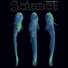
A long-standing goal of biology is to map the behavior of all cells during vertebrate embryogenesis. We developed digital scanned laser light sheet fluorescence microscopy and recorded nuclei localization and movement in entire wild-type and mutant zebrafish embryos over the first 24 hours of development. Multiview in vivo imaging at 1.5 billion voxels per minute provides "digital embryos," that is, comprehensive databases of cell positions, divisions, and migratory tracks. Our analysis of global cell division patterns reveals a maternally defined initial morphodynamic symmetry break, which identifies the embryonic body axis. We further derive a model of germ layer formation and show that the mesendoderm forms from one-third of the embryo’s cells in a single event. Our digital embryos, with 55 million nucleus entries, are provided as a resource.

Although microtubules are key players in many cellular processes, very little is known about their dynamic and mechanical properties in physiological three-dimensional environments. The conventional model of microtubule dynamic instability postulates two dynamic microtubule states, growth and shrinkage. However, several studies have indicated that such a model does not provide a comprehensive quantitative and qualitative description of microtubule behavior. Using three-dimensional laser light-sheet fluorescence microscopy and a three-dimensional sample preparation in spacious Teflon cylinders, we measured microtubule dynamic instability and elasticity in interphase Xenopus laevis egg extracts. Our data are inconsistent with a two-state model of microtubule dynamic instability and favor an extended four-state model with two independent metastable pause states over a three-state model with a single pause state. Moreover, our data on kinetic state transitions rule out a simple GTP cap model as the driving force of microtubule stabilization in egg extracts on timescales of a few seconds or longer. We determined the three-dimensional elastic properties of microtubules as a function of both the contour length and the dynamic state. Our results indicate that pausing microtubules are less flexible than growing microtubules and suggest a growth-speed-dependent persistence length. These data might hint toward mechanisms that enable microtubules to efficiently perform multiple different tasks in the cell and suggest the development of a unified model of microtubule dynamics and microtubule mechanics.

We present an experimental investigation of microtubule dynamic instability in three dimensions, based on laser light-sheet fluorescence microscopy. We introduce three-dimensional (3D) preparation of Xenopus laevis egg extracts in Teflon-based cylinders and provide algorithms for 3D image processing. Our approach gives experimental access to the intrinsic dynamic properties of microtubules and to microtubule population statistics in single asters. We obtain evidence for a stochastic nature of microtubule pausing.

Nud1p, a protein homologous to the mammalian centrosome and midbody component Centriolin, is a component of the budding yeast spindle pole body (SPB), with roles in anchorage of microtubules and regulation of the mitotic exit network during vegetative growth. Here we analyze the function of Nud1p during yeast meiosis. We find that a nud1-2 temperature-sensitive mutant has two meiosis-related defects that reflect genetically distinct functions of Nud1p. First, the mutation affects spore formation due to its late function during spore maturation. Second, and most important, the mutant loses its ability to distinguish between the ages of the four spindle pole bodies, which normally determine which SPB would be preferentially included in the mature spores. This affects the regulation of genome inheritance in starved meiotic cells and leads to the formation of random dyads instead of non-sister dyads under these conditions. Both functions of Nud1p are connected to the ability of Spc72p to bind to the outer plaque and half-bridge (via Kar1p) of the SPB.

Novel technologies are required for three-dimensional cell biology and biophysics. By three-dimensional we refer to experimental conditions that essentially try to avoid hard and flat surfaces and favour unconstrained sample dynamics. We believe that light-sheet-based microscopes are particularly well suited to studies of sensitive three-dimensional biological systems. The application of such instruments can be illustrated with examples from the biophysics of microtubule dynamics and three-dimensional cell cultures. Our experience leads us to suggest that three-dimensional approaches reveal new aspects of a system and enable experiments to be performed in a more physiological and hence clinically more relevant context.

Spindle pole bodies (SPBs) provide a structural basis for genome inheritance and spore formation during meiosis in yeast. Upon carbon source limitation during sporulation, the number of haploid spores formed per cell is reduced. We show that precise spore number control (SNC) fulfills two functions. SNC maximizes the production of spores (1-4) that are formed by a single cell. This is regulated by the concentration of three structural meiotic SPB components, which is dependent on available amounts of carbon source. Using experiments and computer simulation, we show that the molecular mechanism relies on a self-organizing system, which is able to generate particular patterns (different numbers of spores) in dependency on one single stimulus (gradually increasing amounts of SPB constituents). We also show that SNC enhances intratetrad mating, whereby maximal amounts of germinated spores are able to return to a diploid lifestyle without intermediary mitotic division. This is beneficial for the immediate fitness of the population of postmeiotic cells.