Main Menu (Mobile)- Block
Main Menu - Block
Select Publications
View All Publications

We present a method to automatically identify and track nuclei in time-lapse microscopy recordings of entire developing embryos. The method combines deep learning and global optimization. On a mouse dataset, it reconstructs 75.8% of cell lineages spanning 1 h, as compared to 31.8% for the competing method. Our approach improves understanding of where and when cell fate decisions are made in developing embryos, tissues, and organs.
View Publication Page
The mammalian heart is derived from multiple cell lineages; however, our understanding of when and how the diverse cardiac cell types arise is limited. We mapped the origin of the embryonic mouse heart at single-cell resolution using a combination of transcriptomic, imaging, and genetic lineage labeling approaches. This provided a transcriptional and anatomic definition of cardiac progenitor types. Furthermore, it revealed a cardiac progenitor pool that is anatomically and transcriptionally distinct from currently known cardiac progenitors. Besides contributing to cardiomyocytes, these cells also represent the earliest progenitor of the epicardium, a source of trophic factors and cells during cardiac development and injury. This study provides detailed insights into the formation of early cardiac cell types, with particular relevance to the development of cell-based cardiac regenerative therapies.
View Publication Page
Tissue clearing and light-sheet microscopy have a 100-year-plus history, yet these fields have been combined only recently to facilitate novel experiments and measurements in neuroscience. Since tissue-clearing methods were first combined with modernized light-sheet microscopy a decade ago, the performance of both technologies has rapidly improved, broadening their applications. Here, we review the state of the art of tissue-clearing methods and light-sheet microscopy and discuss applications of these techniques in profiling cells and circuits in mice. We examine outstanding challenges and future opportunities for expanding these techniques to achieve brain-wide profiling of cells and circuits in primates and humans. Such integration will help provide a systems-level understanding of the physiology and pathology of our central nervous system.
View Publication Page
State-of-the-art tissue-clearing methods provide subcellular-level optical access to intact tissues from individual organs and even to some entire mammals. When combined with light-sheet microscopy and automated approaches to image analysis, existing tissue-clearing methods can speed up and may reduce the cost of conventional histology by several orders of magnitude. In addition, tissue-clearing chemistry allows whole-organ antibody labelling, which can be applied even to thick human tissues. By combining the most powerful labelling, clearing, imaging and data-analysis tools, scientists are extracting structural and functional cellular and subcellular information on complex mammalian bodies and large human specimens at an accelerated pace. The rapid generation of terabyte-scale imaging data furthermore creates a high demand for efficient computational approaches that tackle challenges in large-scale data analysis and management. In this Review, we discuss how tissue-clearing methods could provide an unbiased, system-level view of mammalian bodies and human specimens and discuss future opportunities for the use of these methods in human neuroscience.
View Publication Page
The ability to visualize and quantitatively measure dynamic biological processes in vivo and at high spatiotemporal resolution is of fundamental importance to experimental investigations in developmental biology. Light-sheet microscopy is particularly well suited to providing such data, since it offers exceptionally high imaging speed and good spatial resolution while minimizing light-induced damage to the specimen. We review core principles and recent advances in light-sheet microscopy, with a focus on concepts and implementations relevant for applications in developmental biology. We discuss how light-sheet microcopy has helped advance our understanding of developmental processes from single-molecule to whole-organism studies, assess the potential for synergies with other state-of-the-art technologies, and introduce methods for computational image and data analysis. Finally, we explore the future trajectory of light-sheet microscopy, discuss key efforts to disseminate new light-sheet technology, and identify exciting opportunities for further advances.
View Publication Page
Animal survival requires a functioning nervous system to develop during embryogenesis. Newborn neurons must assemble into circuits producing activity patterns capable of instructing behaviors. Elucidating how this process is coordinated requires new methods that follow maturation and activity of all cells across a developing circuit. We present an imaging method for comprehensively tracking neuron lineages, movements, molecular identities, and activity in the entire developing zebrafish spinal cord, from neurogenesis until the emergence of patterned activity instructing the earliest spontaneous motor behavior. We found that motoneurons are active first and form local patterned ensembles with neighboring neurons. These ensembles merge, synchronize globally after reaching a threshold size, and finally recruit commissural interneurons to orchestrate the left-right alternating patterns important for locomotion in vertebrates. Individual neurons undergo functional maturation stereotypically based on their birth time and anatomical origin. Our study provides a general strategy for reconstructing how functioning circuits emerge during embryogenesis.
View Publication Page
Whole-brain imaging allows for comprehensive functional mapping of distributed neural pathways, but neuronal perturbation experiments are usually limited to targeting predefined regions or genetically identifiable cell types. To complement whole-brain measures of activity with brain-wide manipulations for testing causal interactions, we introduce a system that uses measuredactivity patterns to guide optical perturbations of any subset of neurons in the same fictively behaving larval zebrafish. First, a light-sheet microscope collects whole-brain data that are rapidly analyzed by a distributed computing system to generate functional brain maps. On the basis of these maps, the experimenter can then optically ablate neurons and image activity changes across the brain. We applied this method to characterize contributions of behaviorally tuned populations to the optomotor response. We extended the system to optogenetically stimulate arbitrary subsets of neurons during whole-brain imaging. These open-source methods enable delineating the contributions of neurons to brain-wide circuit dynamics and behavior in individual animals.
View Publication Page
We describe the implementation and use of an adaptive imaging framework for optimizing spatial resolution and signal strength in a light-sheet microscope. The framework, termed AutoPilot, comprises hardware and software modules for automatically measuring and compensating for mismatches between light-sheet and detection focal planes in living specimens. Our protocol enables researchers to introduce adaptive imaging capabilities in an existing light-sheet microscope or use our SiMView microscope blueprint to set up a new adaptive multiview light-sheet microscope. The protocol describes (i) the mechano-optical implementation of the adaptive imaging hardware, including technical drawings for all custom microscope components; (ii) the algorithms and software library for automated adaptive imaging, including the pseudocode and annotated source code for all software modules; and (iii) the execution of adaptive imaging experiments, as well as the configuration and practical use of the AutoPilot framework. Setup of the adaptive imaging hardware and software takes 1-2 weeks each. Previous experience with light-sheet microscopy and some familiarity with software engineering and building of optical instruments are recommended. Successful implementation of the protocol recovers near diffraction-limited performance in many parts of typical multicellular organisms studied with light-sheet microscopy, such as fruit fly and zebrafish embryos, for which resolution and signal strength are improved two- to fivefold.
View Publication Page
The mouse embryo has long been central to the study of mammalian development; however, elucidating the cell behaviors governing gastrulation and the formation of tissues and organs remains a fundamental challenge. A major obstacle is the lack of live imaging and image analysis technologies capable of systematically following cellular dynamics across the developing embryo. We developed a light-sheet microscope that adapts itself to the dramatic changes in size, shape, and optical properties of the post-implantation mouse embryo and captures its development from gastrulation to early organogenesis at the cellular level. We furthermore developed a computational framework for reconstructing long-term cell tracks, cell divisions, dynamic fate maps, and maps of tissue morphogenesis across the entire embryo. By jointly analyzing cellular dynamics in multiple embryos registered in space and time, we built a dynamic atlas of post-implantation mouse development that, together with our microscopy and computational methods, is provided as a resource.
View Publication Page
Mechanics plays a key role in the development of higher organisms. However, understanding this relationship is complicated by the difficulty of modeling the link between local forces generated at the subcellular level and deformations observed at the tissue and whole-embryo levels. Here we propose an approach first developed for lipid bilayers and cell membranes, in which force-generation by cytoskeletal elements enters a continuum mechanics formulation for the full system in the form of local changes in preferred curvature. This allows us to express and solve the system using only tissue strains. Locations of preferred curvature are simply related to products of gene expression. A solution, in that context, means relaxing the system’s mechanical energy to yield global morphogenetic predictions that accommodate a tendency toward the local preferred curvature, without a need to explicitly model force-generation mechanisms at the molecular level. Our computational framework, which we call SPHARM-MECH, extends a 3D spherical harmonics parameterization known as SPHARM to combine this level of abstraction with a sparse shape representation. The integration of these two principles allows computer simulations to be performed in three dimensions on highly complex shapes, gene expression patterns, and mechanical constraints. We demonstrate our approach by modeling mesoderm invagination in the fruit-fly embryo, where local forces generated by the acto-myosin meshwork in the region of the future mesoderm lead to formation of a ventral tissue fold. The process is accompanied by substantial changes in cell shape and long-range cell movements. Applying SPHARM-MECH to whole-embryo live imaging data acquired with light-sheet microscopy reveals significant correlation between calculated and observed tissue movements. Our analysis predicts the observed cell shape anisotropy on the ventral side of the embryo and suggests an active mechanical role of mesoderm invagination in supporting the onset of germ-band extension.
View Publication Page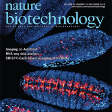
Optimal image quality in light-sheet microscopy requires a perfect overlap between the illuminating light sheet and the focal plane of the detection objective. However, mismatches between the light-sheet and detection planes are common owing to the spatiotemporally varying optical properties of living specimens. Here we present the AutoPilot framework, an automated method for spatiotemporally adaptive imaging that integrates (i) a multi-view light-sheet microscope capable of digitally translating and rotating light-sheet and detection planes in three dimensions and (ii) a computational method that continuously optimizes spatial resolution across the specimen volume in real time. We demonstrate long-term adaptive imaging of entire developing zebrafish (Danio rerio) and Drosophila melanogaster embryos and perform adaptive whole-brain functional imaging in larval zebrafish. Our method improves spatial resolution and signal strength two to five-fold, recovers cellular and sub-cellular structures in many regions that are not resolved by non-adaptive imaging, adapts to spatiotemporal dynamics of genetically encoded fluorescent markers and robustly optimizes imaging performance during large-scale morphogenetic changes in living organisms.
View Publication Page
A custom-built objective lens called the Mesolens allows relatively large biological specimens to be imaged with cellular resolution.
View Publication Page
Animal development is a complex and dynamic process orchestrated by exquisitely timed cell lineage commitment, divisions, migration, and morphological changes at the single-cell level. In the past decade, extensive genetic, stem cell, and genomic studies provided crucial insights into molecular underpinnings and the functional importance of genetic pathways governing various cellular differentiation processes. However, it is still largely unknown how the precise coordination of these pathways is achieved at the whole-organism level and how the highly regulated spatiotemporal choreography of development is established in turn. Here, we discuss the latest technological advances in imaging and single-cell genomics that hold great promise for advancing our understanding of this intricate process. We propose an integrated approach that combines such methods to quantitatively decipher in vivo cellular dynamic behaviors and their underlying molecular mechanisms at the systems level with single-cell, single-molecule resolution.
View Publication Page
We present the Real-time Accurate Cell-shape Extractor (RACE), a high-throughput image analysis framework for automated three-dimensional cell segmentation in large-scale images. RACE is 55–330 times faster and 2–5 times more accurate than state-of-the-art methods. We demonstrate the generality of RACE by extracting cell-shape information from entire Drosophila, zebrafish, and mouse embryos imaged with confocal and light-sheet microscopes. Using RACE, we automatically reconstructed cellular-resolution tissue anisotropy maps across developing Drosophila embryos and quantified differences in cell-shape dynamics in wild-type and mutant embryos. We furthermore integrated RACE with our framework for automated cell lineaging and performed joint segmentation and cell tracking in entire Drosophila embryos. RACE processed these terabyte-sized datasets on a single computer within 1.4 days. RACE is easy to use, as it requires adjustment of only three parameters, takes full advantage of state-of-the-art multi-core processors and graphics cards, and is available as open-source software for Windows, Linux, and Mac OS.
View Publication Page
Imaging fast cellular dynamics across large specimens requires high resolution in all dimensions, high imaging speeds, good physical coverage and low photo-damage. To meet these requirements, we developed isotropic multiview (IsoView) light-sheet microscopy, which rapidly images large specimens via simultaneous light-sheet illumination and fluorescence detection along four orthogonal directions. Combining these four views by means of high-throughput multiview deconvolution yields images with high resolution in all three dimensions. We demonstrate whole-animal functional imaging of Drosophila larvae at a spatial resolution of 1.1-2.5 μm and temporal resolution of 2 Hz for several hours. We also present spatially isotropic whole-brain functional imaging in Danio rerio larvae and spatially isotropic multicolor imaging of fast cellular dynamics across gastrulating Drosophila embryos. Compared with conventional light-sheet microscopy, IsoView microscopy improves spatial resolution at least sevenfold and decreases resolution anisotropy at least threefold. Compared with existing high-resolution light-sheet techniques, IsoView microscopy effectively doubles the penetration depth and provides subsecond temporal resolution for specimens 400-fold larger than could previously be imaged.
View Publication Page
Light-sheet microscopy is a powerful method for imaging the development and function of complex biological systems at high spatiotemporal resolution and over long time scales. Such experiments typically generate terabytes of multidimensional image data, and thus they demand efficient computational solutions for data management, processing and analysis. We present protocols and software to tackle these steps, focusing on the imaging-based study of animal development. Our protocols facilitate (i) high-speed lossless data compression and content-based multiview image fusion optimized for multicore CPU architectures, reducing image data size 30–500-fold; (ii) automated large-scale cell tracking and segmentation; and (iii) visualization, editing and annotation of multiterabyte image data and cell-lineage reconstructions with tens of millions of data points. These software modules are open source. They provide high data throughput using a single computer workstation and are readily applicable to a wide spectrum of biological model systems.
View Publication Page
Understanding how the brain works in tight concert with the rest of the central nervous system (CNS) hinges upon knowledge of coordinated activity patterns across the whole CNS. We present a method for measuring activity in an entire, non-transparent CNS with high spatiotemporal resolution. We combine a light-sheet microscope capable of simultaneous multi-view imaging at volumetric speeds 25-fold faster than the state-of-the-art, a whole-CNS imaging assay for the isolated Drosophila larval CNS and a computational framework for analysing multi-view, whole-CNS calcium imaging data. We image both brain and ventral nerve cord, covering the entire CNS at 2 or 5 Hz with two- or one-photon excitation, respectively. By mapping network activity during fictive behaviours and quantitatively comparing high-resolution whole-CNS activity maps across individuals, we predict functional connections between CNS regions and reveal neurons in the brain that identify type and temporal state of motor programs executed in the ventral nerve cord.
View Publication Page
The nature of nervous system function and development is inherently global, since all components eventually influence one another. Networks communicate through dense synaptic, electric, and modulatory connections and develop through concurrent growth and interlinking of their neurons, processes, glia, and blood vessels. These factors drive the development of techniques capable of imaging neural signaling, anatomy, and developmental processes at ever-larger scales. Here, we discuss the nature of questions benefitting from large-scale imaging techniques and introduce recent applications. We focus on emerging light-sheet microscopy approaches, which are well suited for live imaging of large systems with high spatiotemporal resolution and over long periods of time. We also discuss computational methods suitable for extracting biological information from the resulting system-level image data sets. Together with new tools for reporting and manipulating neuronal activity and gene expression, these techniques promise new insights into the large-scale function and development of neural systems.
View Publication Page
Developments in electrical and optical recording technology are scaling up the size of neuronal populations that can be monitored simultaneously. Light-sheet imaging is rapidly gaining traction as a method for optically interrogating activity in large networks and presents both opportunities and challenges for understanding circuit function.
View Publication Page
The molecular and cellular architecture of the organs in a whole mouse is revealed through optical clearing.
View Publication Page
The origin of chordates has been debated for more than a century, with one key issue being the emergence of the notochord. In vertebrates, the notochord develops by convergence and extension of the chordamesoderm, a population of midline cells of unique molecular identity. We identify a population of mesodermal cells in a developing invertebrate, the marine annelid Platynereis dumerilii, that converges and extends toward the midline and expresses a notochord-specific combination of genes. These cells differentiate into a longitudinal muscle, the axochord, that is positioned between central nervous system and axial blood vessel and secretes a strong collagenous extracellular matrix. Ancestral state reconstruction suggests that contractile mesodermal midline cells existed in bilaterian ancestors. We propose that these cells, via vacuolization and stiffening, gave rise to the chordate notochord.
View Publication Page
The comprehensive reconstruction of cell lineages in complex multicellular organisms is a central goal of developmental biology. We present an open-source computational framework for the segmentation and tracking of cell nuclei with high accuracy and speed. We demonstrate its (i) generality by reconstructing cell lineages in four-dimensional, terabyte-sized image data sets of fruit fly, zebrafish and mouse embryos acquired with three types of fluorescence microscopes, (ii) scalability by analyzing advanced stages of development with up to 20,000 cells per time point at 26,000 cells min(-1) on a single computer workstation and (iii) ease of use by adjusting only two parameters across all data sets and providing visualization and editing tools for efficient data curation. Our approach achieves on average 97.0% linkage accuracy across all species and imaging modalities. Using our system, we performed the first cell lineage reconstruction of early Drosophila melanogaster nervous system development, revealing neuroblast dynamics throughout an entire embryo.
View Publication Page
Morphogenesis, the development of the shape of an organism, is a dynamic process on a multitude of scales, from fast subcellular rearrangements and cell movements to slow structural changes at the whole-organism level. Live-imaging approaches based on light microscopy reveal the intricate dynamics of this process and are thus indispensable for investigating the underlying mechanisms. This Review discusses emerging imaging techniques that can record morphogenesis at temporal scales from seconds to days and at spatial scales from hundreds of nanometers to several millimeters. To unlock their full potential, these methods need to be matched with new computational approaches and physical models that help convert highly complex image data sets into biological insights.
Science Profile: A Research Career in Focus
View Publication Page
Brain function relies on communication between large populations of neurons across multiple brain areas, a full understanding of which would require knowledge of the time-varying activity of all neurons in the central nervous system. Here we use light-sheet microscopy to record activity, reported through the genetically encoded calcium indicator GCaMP5G, from the entire volume of the brain of the larval zebrafish in vivo at 0.8 Hz, capturing more than 80% of all neurons at single-cell resolution. Demonstrating how this technique can be used to reveal functionally defined circuits across the brain, we identify two populations of neurons with correlated activity patterns. One circuit consists of hindbrain neurons functionally coupled to spinal cord neuropil. The other consists of an anatomically symmetric population in the anterior hindbrain, with activity in the left and right halves oscillating in antiphase, on a timescale of 20 s, and coupled to equally slow oscillations in the inferior olive.
View Publication Page
The functional state of a cell is largely determined by the spatiotemporal organization of its proteome. Technologies exist for measuring particular aspects of protein turnover and localization, but comprehensive analysis of protein dynamics across different scales is possible only by combining several methods. Here we describe tandem fluorescent protein timers (tFTs), fusions of two single-color fluorescent proteins that mature with different kinetics, which we use to analyze protein turnover and mobility in living cells. We fuse tFTs to proteins in yeast to study the longevity, segregation and inheritance of cellular components and the mobility of proteins between subcellular compartments; to measure protein degradation kinetics without the need for time-course measurements; and to conduct high-throughput screens for regulators of protein turnover. Our experiments reveal the stable nature and asymmetric inheritance of nuclear pore complexes and identify regulators of N-end rule–mediated protein degradation.
View Publication Page
Live imaging of large biological specimens is fundamentally limited by the short optical penetration depth of light microscopes. To maximize physical coverage, we developed the SiMView technology framework for high-speed in vivo imaging, which records multiple views of the specimen simultaneously. SiMView consists of a light-sheet microscope with four synchronized optical arms, real-time electronics for long-term sCMOS-based image acquisition at 175 million voxels per second, and computational modules for high-throughput image registration, segmentation, tracking and real-time management of the terabytes of multiview data recorded per specimen. We developed one-photon and multiphoton SiMView implementations and recorded cellular dynamics in entire Drosophila melanogaster embryos with 30-s temporal resolution throughout development. We furthermore performed high-resolution long-term imaging of the developing nervous system and followed neuroblast cell lineages in vivo. SiMView data sets provide quantitative morphological information even for fast global processes and enable accurate automated cell tracking in the entire early embryo.
High-resolution movies in the Digital Embryo repository
Nature News: "Fruitfly development, cell by cell" by Lauren Gravitz
Nature Methods Technology Feature: "Faster frames, clearer pictures" by Monya Baker
Andor Insight Awards: Life Sciences Winner

Light sheet microscopy is a versatile imaging technique with a unique combination of capabilities. It provides high imaging speed, high signal-to-noise ratio and low levels of photobleaching and phototoxic effects. These properties are crucial in a wide range of applications in the life sciences, from live imaging of fast dynamic processes in single cells to long-term observation of developmental dynamics in entire large organisms. When combined with tissue clearing methods, light sheet microscopy furthermore allows rapid imaging of large specimens with excellent coverage and high spatial resolution. Even samples up to the size of entire mammalian brains can be efficiently recorded and quantitatively analyzed. Here, we provide an overview of the history of light sheet microscopy, review the development of tissue clearing methods, and discuss recent technical breakthroughs that have the potential to influence the future direction of the field.
View Publication Page
Light sheet-based fluorescence microscopy (LSFM) is emerging as a powerful imaging technique for the life sciences. LSFM provides an exceptionally high imaging speed, high signal-to-noise ratio, low level of photo-bleaching and good optical penetration depth. This unique combination of capabilities makes light sheet-based microscopes highly suitable for live imaging applications. There is an outstanding potential in applying this technology to the quantitative study of embryonic development. Here, we provide an overview of the different basic implementations of LSFM, review recent technical advances in the field and highlight applications in the context of embryonic development. We conclude with a discussion of promising future directions.
View Publication Page
Recording light-microscopy images of large, nontransparent specimens, such as developing multicellular organisms, is complicated by decreased contrast resulting from light scattering. Early zebrafish development can be captured by standard light-sheet microscopy, but new imaging strategies are required to obtain high-quality data of late development or of less transparent organisms. We combined digital scanned laser light-sheet fluorescence microscopy with incoherent structured-illumination microscopy (DSLM-SI) and created structured-illumination patterns with continuously adjustable frequencies. Our method discriminates the specimen-related scattered background from signal fluorescence, thereby removing out-of-focus light and optimizing the contrast of in-focus structures. DSLM-SI provides rapid control of the illumination pattern, exceptional imaging quality, and high imaging speeds. We performed long-term imaging of zebrafish development for 58 h and fast multiple-view imaging of early Drosophila melanogaster development. We reconstructed cell positions over time from the Drosophila DSLM-SI data and created a fly digital embryo.
View Publication Page
During mitosis in Saccharomyces cerevisiae, senescence factors such as extrachromosomal ribosomal DNA circles (ERCs) are retained in the mother cell and excluded from the bud/daughter cell. Shcheprova et al. proposed a model suggesting segregation of ERCs through their association with nuclear pore complexes (NPCs) and retention of preexisting NPCs in the mother cell during mitosis. However, this model is inconsistent with previous data and we demonstrate here that NPCs do efficiently migrate from the mother into the bud. Therefore, binding to NPCs does not seem to explain the retention of ERCs in the mother cell.
View Publication Page
Deleterious mutations inevitably emerge in any evolutionary process and are speculated to decisively influence the structure of the genome. Meiosis, which is thought to play a major role in handling mutations on the population level, recombines chromosomes via non-randomly distributed hot spots for meiotic recombination. In many genomes, various types of genetic elements are distributed in patterns that are currently not well understood. In particular, important (essential) genes are arranged in clusters, which often cannot be explained by a functional relationship of the involved genes. Here we show by computer simulation that essential gene (EG) clustering provides a fitness benefit in handling deleterious mutations in sexual populations with variable levels of inbreeding and outbreeding. We find that recessive lethal mutations enforce a selective pressure towards clustered genome architectures. Our simulations correctly predict (i) the evolution of non-random distributions of meiotic crossovers, (ii) the genome-wide anti-correlation of meiotic crossovers and EG clustering, (iii) the evolution of EG enrichment in pericentromeric regions and (iv) the associated absence of meiotic crossovers (cold centromeres). Our results furthermore predict optimal crossover rates for yeast chromosomes, which match the experimentally determined rates. Using a Saccharomyces cerevisiae conditional mutator strain, we show that haploid lethal phenotypes result predominantly from mutation of single loci and generally do not impair mating, which leads to an accumulation of mutational load following meiosis and mating. We hypothesize that purging of deleterious mutations in essential genes constitutes an important factor driving meiotic crossover. Therefore, the increased robustness of populations to deleterious mutations, which arises from clustered genome architectures, may provide a significant selective force shaping crossover distribution. Our analysis reveals a new aspect of the evolution of genome architectures that complements insights about molecular constraints, such as the interference of pericentromeric crossovers with chromosome segregation.
View Publication Page
The observation of biological processes in their natural in vivo context is a key requirement for quantitative experimental studies in the life sciences. In many instances, it will be crucial to achieve high temporal and spatial resolution over long periods of time without compromising the physiological development of the specimen. Here, we discuss the principles underlying light sheet-based fluorescence microscopes. The most recent implementation DSLM is a tool optimized to deliver quantitative data for entire embryos at high spatio-temporal resolution. We compare DSLM to the two established light microscopy techniques: confocal and two-photon fluorescence microscopy. DSLM provides up to 50 times higher imaging speeds and a 10-100 times higher signal-to-noise ratio, while exposing the specimens to at least three orders of magnitude less light energy than confocal and two-photon fluorescence microscopes. We conclude with a perspective for future development.
View Publication Page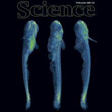
A long-standing goal of biology is to map the behavior of all cells during vertebrate embryogenesis. We developed digital scanned laser light sheet fluorescence microscopy and recorded nuclei localization and movement in entire wild-type and mutant zebrafish embryos over the first 24 hours of development. Multiview in vivo imaging at 1.5 billion voxels per minute provides "digital embryos," that is, comprehensive databases of cell positions, divisions, and migratory tracks. Our analysis of global cell division patterns reveals a maternally defined initial morphodynamic symmetry break, which identifies the embryonic body axis. We further derive a model of germ layer formation and show that the mesendoderm forms from one-third of the embryo’s cells in a single event. Our digital embryos, with 55 million nucleus entries, are provided as a resource.
View Publication Page
We present an experimental investigation of microtubule dynamic instability in three dimensions, based on laser light-sheet fluorescence microscopy. We introduce three-dimensional (3D) preparation of Xenopus laevis egg extracts in Teflon-based cylinders and provide algorithms for 3D image processing. Our approach gives experimental access to the intrinsic dynamic properties of microtubules and to microtubule population statistics in single asters. We obtain evidence for a stochastic nature of microtubule pausing.
View Publication Page
Novel technologies are required for three-dimensional cell biology and biophysics. By three-dimensional we refer to experimental conditions that essentially try to avoid hard and flat surfaces and favour unconstrained sample dynamics. We believe that light-sheet-based microscopes are particularly well suited to studies of sensitive three-dimensional biological systems. The application of such instruments can be illustrated with examples from the biophysics of microtubule dynamics and three-dimensional cell cultures. Our experience leads us to suggest that three-dimensional approaches reveal new aspects of a system and enable experiments to be performed in a more physiological and hence clinically more relevant context.
View Publication Page