Filter
Associated Lab
- Aso Lab (1) Apply Aso Lab filter
- Remove Betzig Lab filter Betzig Lab
- Bock Lab (1) Apply Bock Lab filter
- Clapham Lab (1) Apply Clapham Lab filter
- Fetter Lab (2) Apply Fetter Lab filter
- Harris Lab (7) Apply Harris Lab filter
- Hess Lab (8) Apply Hess Lab filter
- Ji Lab (11) Apply Ji Lab filter
- Lavis Lab (8) Apply Lavis Lab filter
- Lippincott-Schwartz Lab (6) Apply Lippincott-Schwartz Lab filter
- Liu (Zhe) Lab (6) Apply Liu (Zhe) Lab filter
- Magee Lab (2) Apply Magee Lab filter
- Rubin Lab (1) Apply Rubin Lab filter
- Saalfeld Lab (2) Apply Saalfeld Lab filter
- Schreiter Lab (1) Apply Schreiter Lab filter
- Shroff Lab (9) Apply Shroff Lab filter
- Singer Lab (1) Apply Singer Lab filter
- Svoboda Lab (2) Apply Svoboda Lab filter
- Tjian Lab (4) Apply Tjian Lab filter
- Turner Lab (1) Apply Turner Lab filter
Associated Project Team
Publication Date
- 2024 (1) Apply 2024 filter
- 2023 (4) Apply 2023 filter
- 2022 (3) Apply 2022 filter
- 2021 (2) Apply 2021 filter
- 2020 (4) Apply 2020 filter
- 2019 (7) Apply 2019 filter
- 2018 (6) Apply 2018 filter
- 2017 (8) Apply 2017 filter
- 2016 (12) Apply 2016 filter
- 2015 (11) Apply 2015 filter
- 2014 (8) Apply 2014 filter
- 2013 (4) Apply 2013 filter
- 2012 (5) Apply 2012 filter
- 2011 (7) Apply 2011 filter
- 2010 (3) Apply 2010 filter
- 2009 (2) Apply 2009 filter
- 2008 (8) Apply 2008 filter
- 2007 (2) Apply 2007 filter
- 2006 (1) Apply 2006 filter
- 2005 (1) Apply 2005 filter
- 1995 (1) Apply 1995 filter
- 1994 (2) Apply 1994 filter
- 1993 (2) Apply 1993 filter
- 1992 (4) Apply 1992 filter
- 1991 (2) Apply 1991 filter
Type of Publication
110 Publications
Showing 81-90 of 110 resultsWe can resolve multiple discrete features within a focal region of m spatial dimensions by first isolating each on the basis of n >/= 1 unique optical characteristics and then measuring their relative spatial coordinates. The minimum acceptable separation between features depends on the point-spread function in the (m + n)d-dimensional space formed by the spatial coordinates and the optical parameters, whereas the absolute spatial resolution is determined by the accuracy to which the coordinates can be measured. Estimates of each suggest that near-field fluorescence excitation microscopy/spectroscopy with molecular sensitivity and spatial resolution is possible.
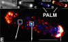
Commentary: Inspired by my earlier work (see below) in single molecule imaging and the isolation of multiple exciton recombination sites within a single probe volume, here I proposed the principle which would eventually lead to PALM. Indeed, all methods of localization microscopy, including PALM, fPALM, PALMIRA, STORM, dSTORM, PAINT, GSDIM, etc. are specific embodiments of the general principle of single molecule isolation and localization I introduced here.
Optical aberrations deteriorate the performance of microscopes. Adaptive optics can be used to improve imaging performance via wavefront shaping. Here, we demonstrate a pupil-segmentation based adaptive optical approach with full-pupil illumination. When implemented in a two-photon fluorescence microscope, it recovers diffraction-limited performance and improves imaging signal and resolution.
Inhomogeneous optical properties of biological samples make it difficult to obtain diffraction-limited resolution in depth. Correcting the sample-induced optical aberrations needs adaptive optics (AO). However, the direct wavefront-sensing approach commonly used in astronomy is not suitable for most biological samples due to their strong scattering of light. We developed an image-based AO approach that is insensitive to sample scattering. By comparing images of the sample taken with different segments of the pupil illuminated, local tilt in the wavefront is measured from image shift. The aberrated wavefront is then obtained either by measuring the local phase directly using interference or with phase reconstruction algorithms similar to those used in astronomical AO. We implemented this pupil-segmentation-based approach in a two-photon fluorescence microscope and demonstrated that diffraction-limited resolution can be recovered from nonbiological and biological samples.
A quantitative understanding of tissue morphogenesis requires description of the movements of individual cells in space and over time. In transparent embryos, such as C. elegans, fluorescently labeled nuclei can be imaged in three-dimensional time-lapse (4D) movies and automatically tracked through early cleavage divisions up to 350 nuclei. A similar analysis of later stages of C. elegans development has been challenging owing to the increased error rates of automated tracking of large numbers of densely packed nuclei. We present Nucleitracker4D, a freely available software solution for tracking nuclei in complex embryos that integrates automated tracking of nuclei in local searches with manual curation. Using these methods, we have been able to track >99% of all nuclei generated in the C. elegans embryo. Our analysis reveals that ventral enclosure of the epidermis is accompanied by complex coordinated migration of the neuronal substrate. We can efficiently track large numbers of migrating nuclei in 4D movies of zebrafish cardiac morphogenesis, suggesting that this approach is generally useful in situations in which the number, packing or dynamics of nuclei present challenges for automated tracking.
Using a descanned, laser-induced guide star and direct wavefront sensing, we demonstrate adaptive correction of complex optical aberrations at high numerical aperture (NA) and a 14-ms update rate. This correction permits us to compensate for the rapid spatial variation in aberration often encountered in biological specimens and to recover diffraction-limited imaging over large volumes (>240 mm per side). We applied this to image fine neuronal processes and subcellular dynamics within the zebrafish brain.
A key challenge when imaging living cells is how to noninvasively extract the most spatiotemporal information possible. Unlike popular wide-field and confocal methods, plane-illumination microscopy limits excitation to the information-rich vicinity of the focal plane, providing effective optical sectioning and high speed while minimizing out-of-focus background and premature photobleaching. Here we used scanned Bessel beams in conjunction with structured illumination and/or two-photon excitation to create thinner light sheets (<0.5 μm) better suited to three-dimensional (3D) subcellular imaging. As demonstrated by imaging the dynamics of mitochondria, filopodia, membrane ruffles, intracellular vesicles and mitotic chromosomes in live cells, the microscope currently offers 3D isotropic resolution down to \~{}0.3 μm, speeds up to nearly 200 image planes per second and the ability to noninvasively acquire hundreds of 3D data volumes from single living cells encompassing tens of thousands of image frames.

A key challenge when imaging living cells is how to noninvasively extract the most spatiotemporal information possible. Unlike popular wide-field and confocal methods, plane-illumination microscopy limits excitation to the information-rich vicinity of the focal plane, providing effective optical sectioning and high speed while minimizing out-of-focus background and premature photobleaching. Here we used scanned Bessel beams in conjunction with structured illumination and/or two-photon excitation to create thinner light sheets (<0.5 μm) better suited to three-dimensional (3D) subcellular imaging. As demonstrated by imaging the dynamics of mitochondria, filopodia, membrane ruffles, intracellular vesicles and mitotic chromosomes in live cells, the microscope currently offers 3D isotropic resolution down to \~{}0.3 μm, speeds up to nearly 200 image planes per second and the ability to noninvasively acquire hundreds of 3D data volumes from single living cells encompassing tens of thousands of image frames.
Commentary: Plane illumination microscopy has proven to be a powerful tool for studying multicellular organisms and their development at single cell resolution. However, the light sheets employed are usually too thick to provide much benefit for imaging organelles within single cultured cells. Here we introduce the use of scanned Bessel beams to create much thinner light sheets better suited to long-term dynamic live cell imaging. Such light sheets not only minimize photobleaching and phototoxicity at the sub-cellular level, but also provide axial resolution enhancement, yielding isotropic three dimensional spatial resolution. Numerous movies are provided to demonstrate the wealth of 4D information (x,y,x,t) that can be obtained from single living cells by the method. Besides providing an attractive alternative to spinning disk, AOD-driven, or line scan confocal microscopes for high speed live cell imaging, the Bessel microscope might serve as a valuable platform for superresolution microscopy (PALM, structured Illumination, or RESOLFT), since confinement of the excitation to the focal plane makes far better use of the limited fluorescence photon budget than does the traditional epi-illumination configuration.
The presumptive altered dynamics of transient molecular interactions in vivo contributing to neurodegenerative diseases have remained elusive. Here, using single-molecule localization microscopy, we show that disease-inducing Huntingtin (mHtt) protein fragments display three distinct dynamic states in living cells - 1) fast diffusion, 2) dynamic clustering and 3) stable aggregation. Large, stable aggregates of mHtt exclude chromatin and form 'sticky' decoy traps that impede target search processes of key regulators involved in neurological disorders. Functional domain mapping based on super-resolution imaging reveals an unexpected role of aromatic amino acids in promoting protein-mHtt aggregate interactions. Genome-wide expression analysis and numerical simulation experiments suggest mHtt aggregates reduce transcription factor target site sampling frequency and impair critical gene expression programs in striatal neurons. Together, our results provide insights into how mHtt dynamically forms aggregates and disrupts the finely-balanced gene control mechanisms in neuronal cells.
RNA granules have been likened to liquid droplets whose dynamics depend on the controlled dissolution and condensation of internal components. The molecules and reactions that drive these dynamics in vivo are not well understood. In this study, we present evidence that a group of intrinsically disordered, serine-rich proteins regulate the dynamics of P granules in C. elegans embryos. The MEG (maternal-effect germline defective) proteins are germ plasm components that are required redundantly for fertility. We demonstrate that MEG-1 and MEG-3 are substrates of the kinase MBK-2/DYRK and the phosphatase PP2A(PPTR-½). Phosphorylation of the MEGs promotes granule disassembly and dephosphorylation promotes granule assembly. Using lattice light sheet microscopy on live embryos, we show that GFP-tagged MEG-3 localizes to a dynamic domain that surrounds and penetrates each granule. We conclude that, despite their liquid-like behavior, P granules are non-homogeneous structures whose assembly in embryos is regulated by phosphorylation.
The Escherichia coli chemotaxis network is a model system for biological signal processing. In E. coli, transmembrane receptors responsible for signal transduction assemble into large clusters containing several thousand proteins. These sensory clusters have been observed at cell poles and future division sites. Despite extensive study, it remains unclear how chemotaxis clusters form, what controls cluster size and density, and how the cellular location of clusters is robustly maintained in growing and dividing cells. Here, we use photoactivated localization microscopy (PALM) to map the cellular locations of three proteins central to bacterial chemotaxis (the Tar receptor, CheY, and CheW) with a precision of 15 nm. We find that cluster sizes are approximately exponentially distributed, with no characteristic cluster size. One-third of Tar receptors are part of smaller lateral clusters and not of the large polar clusters. Analysis of the relative cellular locations of 1.1 million individual proteins (from 326 cells) suggests that clusters form via stochastic self-assembly. The super-resolution PALM maps of E. coli receptors support the notion that stochastic self-assembly can create and maintain approximately periodic structures in biological membranes, without direct cytoskeletal involvement or active transport.
Commentary: Our goal as tool developers is to invent methods capable of uncovering new biological insights unobtainable by pre-existing technologies. A terrific example is given by this paper, where grad students Derek Greenfield and Ann McEvoy in Jan Liphardt’s group at Berkeley used our PALM to image the size and position distributions of chemotaxis proteins in E. Coli with unprecedented precision and sensitivity. Their analysis revealed that the cluster sizes follow a stretched exponential distribution, and the density of clusters is highest furthest away from the largest (e.g., polar) clusters. Both observations support a model for passive self-assembly rather than active cytoskeletal assembly of the chemotaxis network.