Filter
Associated Lab
- Ahrens Lab (5) Apply Ahrens Lab filter
- Branson Lab (3) Apply Branson Lab filter
- Card Lab (1) Apply Card Lab filter
- Cardona Lab (1) Apply Cardona Lab filter
- Druckmann Lab (1) Apply Druckmann Lab filter
- Freeman Lab (4) Apply Freeman Lab filter
- Funke Lab (2) Apply Funke Lab filter
- Harris Lab (1) Apply Harris Lab filter
- Ji Lab (1) Apply Ji Lab filter
- Remove Keller Lab filter Keller Lab
- Lavis Lab (2) Apply Lavis Lab filter
- Liu (Zhe) Lab (1) Apply Liu (Zhe) Lab filter
- Looger Lab (5) Apply Looger Lab filter
- Pavlopoulos Lab (1) Apply Pavlopoulos Lab filter
- Tillberg Lab (1) Apply Tillberg Lab filter
- Turaga Lab (1) Apply Turaga Lab filter
Associated Project Team
Publication Date
- 2024 (1) Apply 2024 filter
- 2023 (1) Apply 2023 filter
- 2022 (1) Apply 2022 filter
- 2021 (2) Apply 2021 filter
- 2020 (5) Apply 2020 filter
- 2019 (6) Apply 2019 filter
- 2018 (6) Apply 2018 filter
- 2017 (2) Apply 2017 filter
- 2016 (6) Apply 2016 filter
- 2015 (8) Apply 2015 filter
- 2014 (8) Apply 2014 filter
- 2013 (10) Apply 2013 filter
- 2012 (3) Apply 2012 filter
- 2011 (4) Apply 2011 filter
- 2010 (4) Apply 2010 filter
- 2009 (1) Apply 2009 filter
- 2008 (3) Apply 2008 filter
- 2007 (1) Apply 2007 filter
- 2006 (2) Apply 2006 filter
- 2005 (1) Apply 2005 filter
Type of Publication
75 Publications
Showing 31-40 of 75 results
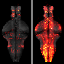
Glucose is arguably the most important molecule in metabolism, and its dysregulation underlies diabetes. We describe a family of single-wavelength genetically encoded glucose sensors with a high signal-to-noise ratio, fast kinetics, and affinities varying over four orders of magnitude (1 μM to 10 mM). The sensors allow mechanistic characterization of glucose transporters expressed in cultured cells with high spatial and temporal resolution. Imaging of neuron/glia co-cultures revealed ∼3-fold faster glucose changes in astrocytes. In larval Drosophila central nervous system explants, intracellular neuronal glucose fluxes suggested a rostro-caudal transport pathway in the ventral nerve cord neuropil. In zebrafish, expected glucose-related physiological sequelae of insulin and epinephrine treatments were directly visualized. Additionally, spontaneous muscle twitches induced glucose uptake in muscle, and sensory and pharmacological perturbations produced large changes in the brain. These sensors will enable rapid, high-resolution imaging of glucose influx, efflux, and metabolism in behaving animals.

Novel technologies are required for three-dimensional cell biology and biophysics. By three-dimensional we refer to experimental conditions that essentially try to avoid hard and flat surfaces and favour unconstrained sample dynamics. We believe that light-sheet-based microscopes are particularly well suited to studies of sensitive three-dimensional biological systems. The application of such instruments can be illustrated with examples from the biophysics of microtubule dynamics and three-dimensional cell cultures. Our experience leads us to suggest that three-dimensional approaches reveal new aspects of a system and enable experiments to be performed in a more physiological and hence clinically more relevant context.
Light sheet-based fluorescence microscopy (LSFM) is emerging as a powerful imaging technique for the life sciences. LSFM provides an exceptionally high imaging speed, high signal-to-noise ratio, low level of photo-bleaching, and good optical penetration depth. This unique combination of capabilities makes light sheet-based microscopes highly suitable for live imaging applications. Here, we provide an overview of light sheet-based microscopy assays for in vitro and in vivo imaging of biological samples, including cell extracts, soft gels, and large multicellular organisms. We furthermore describe computational tools for basic image processing and data inspection.

Light sheet microscopy is a versatile imaging technique with a unique combination of capabilities. It provides high imaging speed, high signal-to-noise ratio and low levels of photobleaching and phototoxic effects. These properties are crucial in a wide range of applications in the life sciences, from live imaging of fast dynamic processes in single cells to long-term observation of developmental dynamics in entire large organisms. When combined with tissue clearing methods, light sheet microscopy furthermore allows rapid imaging of large specimens with excellent coverage and high spatial resolution. Even samples up to the size of entire mammalian brains can be efficiently recorded and quantitatively analyzed. Here, we provide an overview of the history of light sheet microscopy, review the development of tissue clearing methods, and discuss recent technical breakthroughs that have the potential to influence the future direction of the field.
The fruit fly is an excellent model system for investigating the sequence of epithelial tissue invaginations constituting the process of gastrulation. By combining recent advancements in light sheet fluorescence microscopy (LSFM) and image processing, the three-dimensional fly embryo morphology and relevant gene expression patterns can be accurately recorded throughout the entire process of embryogenesis. LSFM provides exceptionally high imaging speed, high signal-to-noise ratio, low level of photoinduced damage, and good optical penetration depth. This powerful combination of capabilities makes LSFM particularly suitable for live imaging of the fly embryo.The resulting high-information-content image data are subsequently processed to obtain the outlines of cells and cell nuclei, as well as the geometry of the whole embryo tissue by image segmentation. Furthermore, morphodynamics information is extracted by computationally tracking objects in the image. Towards that goal we describe the successful implementation of a fast fitting strategy of Gaussian mixture models.The data obtained by image processing is well-suited for hypothesis testing of the detailed biomechanics of the gastrulating embryo. Typically this involves constructing computational mechanics models that consist of an objective function providing an estimate of strain energy for a given morphological configuration of the tissue, and a numerical minimization mechanism of this energy, achieved by varying morphological parameters.In this chapter, we provide an overview of in vivo imaging of fruit fly embryos using LSFM, computational tools suitable for processing the resulting images, and examples of computational biomechanical simulations of fly embryo gastrulation.
The processing of sensory input and the generation of behavior involves large networks of neurons, which necessitates new technology for recording from many neurons in behaving animals. In the larval zebrafish, light-sheet microscopy can be used to record the activity of almost all neurons in the brain simultaneously at single-cell resolution. Existing implementations, however, cannot be combined with visually driven behavior because the light sheet scans over the eye, interfering with presentation of controlled visual stimuli. Here we describe a system that overcomes the confounding eye stimulation through the use of two light sheets and combines whole-brain light-sheet imaging with virtual reality for fictively behaving larval zebrafish.
Developments in electrical and optical recording technology are scaling up the size of neuronal populations that can be monitored simultaneously. Light-sheet imaging is rapidly gaining traction as a method for optically interrogating activity in large networks and presents both opportunities and challenges for understanding circuit function.

The ability to visualize and quantitatively measure dynamic biological processes in vivo and at high spatiotemporal resolution is of fundamental importance to experimental investigations in developmental biology. Light-sheet microscopy is particularly well suited to providing such data, since it offers exceptionally high imaging speed and good spatial resolution while minimizing light-induced damage to the specimen. We review core principles and recent advances in light-sheet microscopy, with a focus on concepts and implementations relevant for applications in developmental biology. We discuss how light-sheet microcopy has helped advance our understanding of developmental processes from single-molecule to whole-organism studies, assess the potential for synergies with other state-of-the-art technologies, and introduce methods for computational image and data analysis. Finally, we explore the future trajectory of light-sheet microscopy, discuss key efforts to disseminate new light-sheet technology, and identify exciting opportunities for further advances.
This protocol describes how to observe gastrulation in living mouse embryos by using light-sheet microscopy and computational tools to analyze the resulting image data at the single-cell level. We describe a series of techniques needed to image the embryos under physiological conditions, including how to hold mouse embryos without agarose embedding, how to transfer embryos without air exposure and how to construct environmental chambers for live imaging by digital scanned light-sheet microscopy (DSLM). Computational tools include manual and semiautomatic tracking programs that are developed for analyzing the large 4D data sets acquired with this system. Note that this protocol does not include details of how to build the light-sheet microscope itself. Time-lapse imaging ends within 12 h, with subsequent tracking analysis requiring 3-6 d. Other than some mouse-handling skills, this protocol requires no advanced skills or knowledge. Light-sheet microscopes are becoming more widely available, and thus the techniques outlined in this paper should be helpful for investigating mouse embryogenesis.

In vivo imaging applications typically require carefully balancing conflicting parameters. Often it is necessary to achieve high imaging speed, low photo-bleaching, and photo-toxicity, good three-dimensional resolution, high signal-to-noise ratio, and excellent physical coverage at the same time. Light-sheet microscopy provides good performance in all of these categories, and is thus emerging as a particularly powerful live imaging method for the life sciences. We see an outstanding potential for applying light-sheet microscopy to the study of development and function of the early nervous system in vertebrates and higher invertebrates. Here, we review state-of-the-art approaches to live imaging of early development, and show how the unique capabilities of light-sheet microscopy can further advance our understanding of the development and function of the nervous system. We discuss key considerations in the design of light-sheet microscopy experiments, including sample preparation and fluorescent marker strategies, and provide an outlook for future directions in the field.