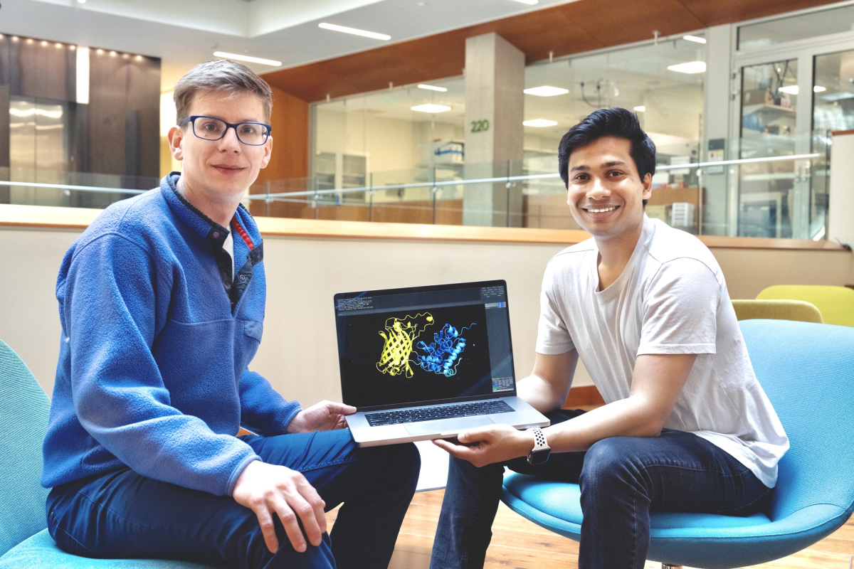
The best part about developing a state-of-the-art neurotransmitter sensor, according to Kasper Podgorski and Abhi Aggarwal, isn’t the recently published paper or the emails from grateful researchers around the world.
Instead, it’s the close collaboration and friendship forged during long discussions in Bob’s pub and late nights at the lab bench that the two scientists will treasure most about developing the glutamate indicator iGluSnFR3.
“This really is a story about how fun it was for us to work at Janelia,” says Podgorski, who was a Janelia Fellow from 2015 to 2021 and is now a senior scientist at the Allen Institute. “It’s about us spending all our free time in the lab working on something we were very passionate about and that we will remember for the rest of our lives as a fun project.”
The collaboration started when Aggarwal came to Janelia in the summer of 2019 as a Janelia Undergraduate Scholar. Podgorski, a microscopist, had recently embarked on a project to devise a genetically encoded indicator to measure the neurotransmitter glutamate. He thought it would be fun to tackle it together with Aggarwal over the summer.
Glutamate is one of the most important and abundant neurotransmitters in the brain, playing a crucial role in learning, memory, and information processing. It helps transmit information between neurons by generating an electrical signal in a receiving neuron that is then sent out to other neurons in the brain. Being able to see and measure glutamate could help scientists understand how neurons communicate and transform inputs into outputs to perform computations.
“My whole career has been driven by trying to find out how neurons compute. To me, that’s a huge gap in neuroscience, and one of the foremost questions that we as neuroscientists should be trying to understand,” Podgorski says. “And the best way forward to understand neuronal computation is to make an indicator that works for synaptic glutamate.”
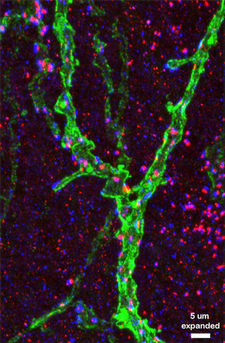
High speed video of cultured neurons expressing iGluSnFR3 (100 Hz, measured on a simple widefield microscope). Neurons have been silenced with TTX, blocking evoked glutamate release. The flashes are iGluSnFR3 responding to spontaneous release of individual vesicles, on average containing just 500 molecules of glutamate. Credit: Aggarwal et al.
Right place, right time
Glutamate indicators, which cause neurons to light up when they sense glutamate, had been developed in 2013 and enhanced in 2018 by former Janelia Group Leader Loren Looger and Senior Scientist Jonathan Marvin. But these earlier sensors had not yet been optimized for measuring glutamate entering a synapse – the space where two communicating neurons meet. They also illuminated glutamate in other parts of the cell, like the shafts of dendrites. Podgorski wanted to develop a sensor that lit up only the glutamate entering a synapse, which could help researchers home in on the input coming into the cell.
Podgorski didn’t have much experience in protein engineering, but luckily he was just down the hall from Looger, Marvin, and Janelia Group Leader Eric Schreiter. They helped Podgorski learn protein engineering and gave him lab space for the project.
“Nowhere else but at Janelia do you see a junior scientist leading a microscopy lab embarking on large-scale protein engineering,” says Podgorski.

Aggarwal joined Podgorski on the project when he showed up in the summer of 2019 after graduating from the University of Alberta, where he had gained experience in developing protein-based sensors.
“When I started, I didn’t know that this is what we were going to do,” says Aggarwal, who is now a research associate at the Allen Institute. “I thought I would join and stay for four months, but I ended up staying for four years.”
The pair worked with Janelia’s GENIE project team and collaborating labs to test different glutamate indicator variants, eventually focusing on iGluSnFR3. Currently the fastest and brightest indicator available for measuring glutamate as it enters a neuron’s dendrites, the sensor is detailed in a new paper in Nature Methods.
The increased sensitivity and faster speed of iGluSnFR3, first released in 2022 and now used by dozens of labs around the world, allows scientists to see the release of glutamate from individual synaptic vesicles at hundreds of synapses at the same time.
“It enables us to see what individual synapses are doing, as opposed to the general milieu inside the brain,” Podgorski says. “By measuring the inputs to the cell and its output, we can understand what the function is that the neuron performs, in the same way that a mathematical function turns inputs into outputs.”
Podgorski and Aggarwal, now both part of the Optophysiology program at the Allen Institute for Neural Dynamics, are continuing their collaboration with the GENIE project team to develop glutamate sensors that are even faster and more sensitive. But it was the development of iGluSnFR3 that will always hold a special place in their hearts.
“It really brought us together and we’ve been great friends ever since,” Podgorski says.
###
Citation:
Abhi Aggarwal, Rui Liu, Yang Chen, Amelia J. Ralowicz, Samuel J. Bergerson, Filip Tomaska, Boaz Mohar, Timothy L. Hanson, Jeremy P. Hasseman, Daniel Reep, Getahun Tsegaye, Pantong Yao, Xiang Ji, Marinus Kloos, Deepika Walpita, Ronak Patel, Manuel A. Mohr, Paul W. Tillberg, The GENIE Project Team, Loren L. Looger, Jonathan S. Marvin, Michael B. Hoppa, Arthur Konnerth, David Kleinfeld, Eric R. Schreiter & Kaspar Podgorski. “Glutamate indicators with improved activation kinetics and localization for imaging synaptic transmission.” Nature Methods. Published May 4, 2023. doi: 10.1038/s41592-023-01863-6
Media Contacts
Nanci Bompey


