Filter
Associated Lab
- Ahrens Lab (5) Apply Ahrens Lab filter
- Branson Lab (3) Apply Branson Lab filter
- Card Lab (1) Apply Card Lab filter
- Cardona Lab (1) Apply Cardona Lab filter
- Druckmann Lab (1) Apply Druckmann Lab filter
- Freeman Lab (4) Apply Freeman Lab filter
- Funke Lab (2) Apply Funke Lab filter
- Harris Lab (1) Apply Harris Lab filter
- Ji Lab (1) Apply Ji Lab filter
- Remove Keller Lab filter Keller Lab
- Lavis Lab (2) Apply Lavis Lab filter
- Liu (Zhe) Lab (1) Apply Liu (Zhe) Lab filter
- Looger Lab (5) Apply Looger Lab filter
- Pavlopoulos Lab (1) Apply Pavlopoulos Lab filter
- Tillberg Lab (1) Apply Tillberg Lab filter
- Turaga Lab (1) Apply Turaga Lab filter
Associated Project Team
Publication Date
- 2024 (1) Apply 2024 filter
- 2023 (1) Apply 2023 filter
- 2022 (1) Apply 2022 filter
- 2021 (2) Apply 2021 filter
- 2020 (5) Apply 2020 filter
- 2019 (6) Apply 2019 filter
- 2018 (6) Apply 2018 filter
- 2017 (2) Apply 2017 filter
- 2016 (6) Apply 2016 filter
- 2015 (8) Apply 2015 filter
- 2014 (8) Apply 2014 filter
- 2013 (10) Apply 2013 filter
- 2012 (3) Apply 2012 filter
- 2011 (4) Apply 2011 filter
- 2010 (4) Apply 2010 filter
- 2009 (1) Apply 2009 filter
- 2008 (3) Apply 2008 filter
- 2007 (1) Apply 2007 filter
- 2006 (2) Apply 2006 filter
- 2005 (1) Apply 2005 filter
Type of Publication
75 Publications
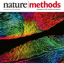
Showing 21-30 of 75 resultsThe comprehensive reconstruction of cell lineages in complex multicellular organisms is a central goal of developmental biology. We present an open-source computational framework for the segmentation and tracking of cell nuclei with high accuracy and speed. We demonstrate its (i) generality by reconstructing cell lineages in four-dimensional, terabyte-sized image data sets of fruit fly, zebrafish and mouse embryos acquired with three types of fluorescence microscopes, (ii) scalability by analyzing advanced stages of development with up to 20,000 cells per time point at 26,000 cells min(-1) on a single computer workstation and (iii) ease of use by adjusting only two parameters across all data sets and providing visualization and editing tools for efficient data curation. Our approach achieves on average 97.0% linkage accuracy across all species and imaging modalities. Using our system, we performed the first cell lineage reconstruction of early Drosophila melanogaster nervous system development, revealing neuroblast dynamics throughout an entire embryo.
Recording light-microscopy images of large, nontransparent specimens, such as developing multicellular organisms, is complicated by decreased contrast resulting from light scattering. Early zebrafish development can be captured by standard light-sheet microscopy, but new imaging strategies are required to obtain high-quality data of late development or of less transparent organisms. We combined digital scanned laser light-sheet fluorescence microscopy with incoherent structured-illumination microscopy (DSLM-SI) and created structured-illumination patterns with continuously adjustable frequencies. Our method discriminates the specimen-related scattered background from signal fluorescence, thereby removing out-of-focus light and optimizing the contrast of in-focus structures. DSLM-SI provides rapid control of the illumination pattern, exceptional imaging quality, and high imaging speeds. We performed long-term imaging of zebrafish development for 58 h and fast multiple-view imaging of early Drosophila melanogaster development. We reconstructed cell positions over time from the Drosophila DSLM-SI data and created a fly digital embryo.
Histone post-translational modifications are key gene expression regulators, but their rapid dynamics during development remain difficult to capture. We applied a Fab-based live endogenous modification labeling technique to monitor the changes in histone modification levels during zygotic genome activation (ZGA) in living zebrafish embryos. Among various histone modifications, H3 Lys27 acetylation (H3K27ac) exhibited most drastic changes, accumulating in two nuclear foci in the 64- to 1k-cell-stage embryos. The elongating form of RNA polymerase II, which is phosphorylated at Ser2 in heptad repeats within the C-terminal domain (RNAP2 Ser2ph), and miR-430 transcripts were also concentrated in foci closely associated with H3K27ac. When treated with α-amanitin to inhibit transcription or JQ-1 to inhibit binding of acetyl-reader proteins, H3K27ac foci still appeared but RNAP2 Ser2ph and miR-430 morpholino were not concentrated in foci, suggesting that H3K27ac precedes active transcription during ZGA. We anticipate that the method presented here could be applied to a variety of developmental processes in any model and non-model organisms.
A custom-built objective lens called the Mesolens allows relatively large biological specimens to be imaged with cellular resolution.
Morphogenesis, the development of the shape of an organism, is a dynamic process on a multitude of scales, from fast subcellular rearrangements and cell movements to slow structural changes at the whole-organism level. Live-imaging approaches based on light microscopy reveal the intricate dynamics of this process and are thus indispensable for investigating the underlying mechanisms. This Review discusses emerging imaging techniques that can record morphogenesis at temporal scales from seconds to days and at spatial scales from hundreds of nanometers to several millimeters. To unlock their full potential, these methods need to be matched with new computational approaches and physical models that help convert highly complex image data sets into biological insights.
The mouse embryo has long been central to the study of mammalian development; however, elucidating the cell behaviors governing gastrulation and the formation of tissues and organs remains a fundamental challenge. A major obstacle is the lack of live imaging and image analysis technologies capable of systematically following cellular dynamics across the developing embryo. We developed a light-sheet microscope that adapts itself to the dramatic changes in size, shape, and optical properties of the post-implantation mouse embryo and captures its development from gastrulation to early organogenesis at the cellular level. We furthermore developed a computational framework for reconstructing long-term cell tracks, cell divisions, dynamic fate maps, and maps of tissue morphogenesis across the entire embryo. By jointly analyzing cellular dynamics in multiple embryos registered in space and time, we built a dynamic atlas of post-implantation mouse development that, together with our microscopy and computational methods, is provided as a resource.
Glucose is arguably the most important molecule in metabolism, and its mismanagement underlies diseases of vast societal import, most notably diabetes. Although glucose-related metabolism has been the subject of intense study for over a century, tools to track glucose in living organisms with high spatio-temporal resolution are lacking. We describe the engineering of a family of genetically encoded glucose sensors with high signal-to-noise ratio, fast kinetics and affinities varying over four orders of magnitude (1 µM to 10 mM). The sensors allow rigorous mechanistic characterization of glucose transporters expressed in cultured cells with high spatial and temporal resolution. Imaging of neuron/glia co-cultures revealed ∼3-fold higher glucose changes in astrocytes versus neurons. In larval Drosophila central nervous system explants, imaging of intracellular neuronal glucose suggested a novel rostro-caudal transport pathway in the ventral nerve cord neuropil, with paradoxically slower uptake into the peripheral cell bodies and brain lobes. In living zebrafish, expected glucose-related physiological sequelae of insulin and epinephrine treatments were directly visualized in real time. Additionally, spontaneous muscle twitches induced glucose uptake in muscle, and sensory- and pharmacological perturbations gave rise to large but enigmatic changes in the brain. These sensors will enable myriad experiments, most notably rapid, high-resolution imaging of glucose influx, efflux, and metabolism in behaving animals.
View Publication PageThe zebrafish Danio rerio has emerged as a powerful vertebrate model system that lends itself particularly well to quantitative investigations with live imaging approaches, owing to its exceptionally high optical clarity in embryonic and larval stages. Recent advances in light microscopy technology enable comprehensive analyses of cellular dynamics during zebrafish embryonic development, systematic mapping of gene expression dynamics, quantitative reconstruction of mutant phenotypes and the system-level biophysical study of morphogenesis. Despite these technical breakthroughs, it remains challenging to design and implement experiments for in vivo long-term imaging at high spatio-temporal resolution. This article discusses the fundamental challenges in zebrafish long-term live imaging, provides experimental protocols and highlights key properties and capabilities of advanced fluorescence microscopes. The article focuses in particular on experimental assays based on light sheet-based fluorescence microscopy, an emerging imaging technology that achieves exceptionally high imaging speeds and excellent signal-to-noise ratios, while minimizing light-induced damage to the specimen. This unique combination of capabilities makes light sheet microscopy an indispensable tool for the in vivo long-term imaging of large developing organisms.
In the field of biomedical imaging analysis on single-cell level, reliable and fast segmentation of the cell nuclei from the background on three-dimensional images is highly needed for the further analysis. In this work we propose an interactive cell segmentation toolkit that first establishes a set of exemplar regions from user input through an easy and intuitive interface in both 2D and 3D in real-time, then
extracts the shape and intensity features from those exemplars. Based on a local contrast-constrained region growing scheme, each connected component in the whole image would be filtered by the features from exemplars, forming an “exemplar-matching” group which passed the filtering and would be part of the final segmentation result, and a “non-exemplar-matching” group in which components
would be further segmented by the gradient vector field based algorithm. The results of the filtering process are visualized back to the user in near real-time, thus enhancing the experience in exemplar selecting and parameter tuning. The toolkit is distributed as a plugin within the open source Vaa3D system (http://vaa3d.org).

