Main Menu (Mobile)- Block

Main Menu - Block

Molecular Tools and Imaging

area_landing_page_panel-header | block

Molecular Tools and Imaging

Inventing novel reagents and technologies that push the boundaries of biological discovery.

area_landing_page_panel-summary | block

Summary

Light bulb icon

Janelias tool-builders are independent experts in a range of physical, chemical, and biological disciplines who develop creative solutions for problems in biology.

Microscope icon

We develop microscopes that allow imaging of biological systems with unprecedented resolution and depth as well as new imaging agents that enable measurement of phenomena inside cells or animals. 

Globe icon

We also freely share our tools – a hallmark of the Janelia ethos – and the microscopes, dyes, and sensors developed at Janelia are being used in labs around the world.

As Janelia moves into new research areas, labs in the Molecular Tools and Imaging program will continue to invent novel reagents and technologies that push the boundaries of biological discovery.

area_landing_page_panel-news | block

News

NEWS

09/26/19 | How Neural Circuits Form in a Developing Embryo: A new imaging method follows young neurons in a developing embryo as they progress from a messy jumble of cells into a coordinated control center. The approach lets scientists track development and emerging cell function simultaneously across entire circuits.

ANNOUNCEMENT

09/03/19 | Please welcome our newest group leader, Abraham Beyene! He’ll join us at Janelia soon. 

Load More
area_landing_page_panel-spotlight | block
area_landing_page_panel-slideshow | block

Our Work

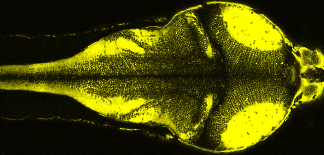
Fluorescent dye shows which neurons are active in a zebrafish

Voltron fish

Janelia scientists develop new ways to track neural activity. One tool, called Voltron, pairs a multipart voltage-sensitive protein with bright and stable dyes, allowing researchers to track neuron activity in living animals more precisely and for far longer time periods than was once possible.

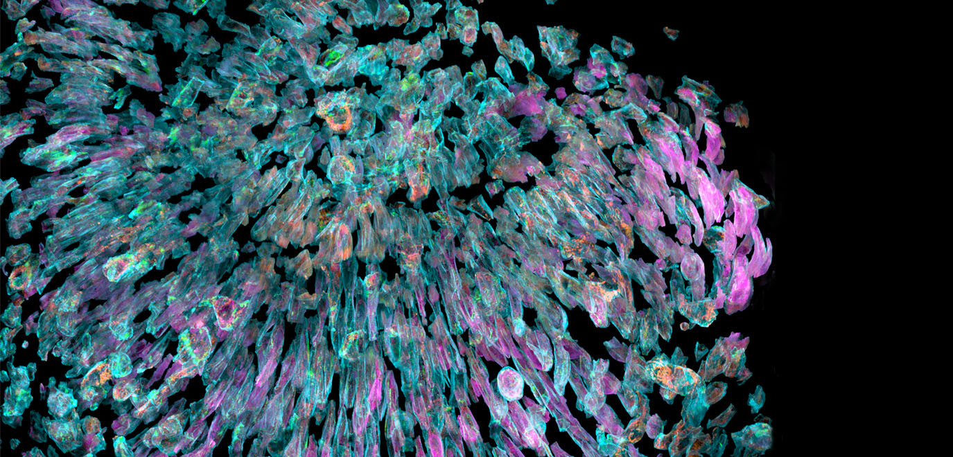
Organelles inside zebrafish eye cells

Organelle dynamics across the zebrafish eye

Merging two imaging technologies – lattice light-sheet microscopy and adaptive optics – reveals the most detailed picture yet of subcellular dynamics in multicellular organisms.

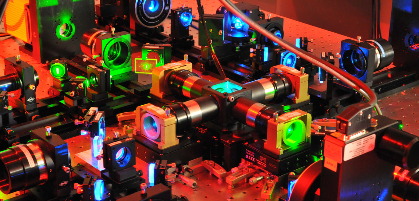
Machinery inside isoview light sheet microscope

IsoView light-sheet microscope

This technology from the Keller Lab allows researchers to visualize whole-brain activity and entire developing embryos in real time at subcellular resolution.

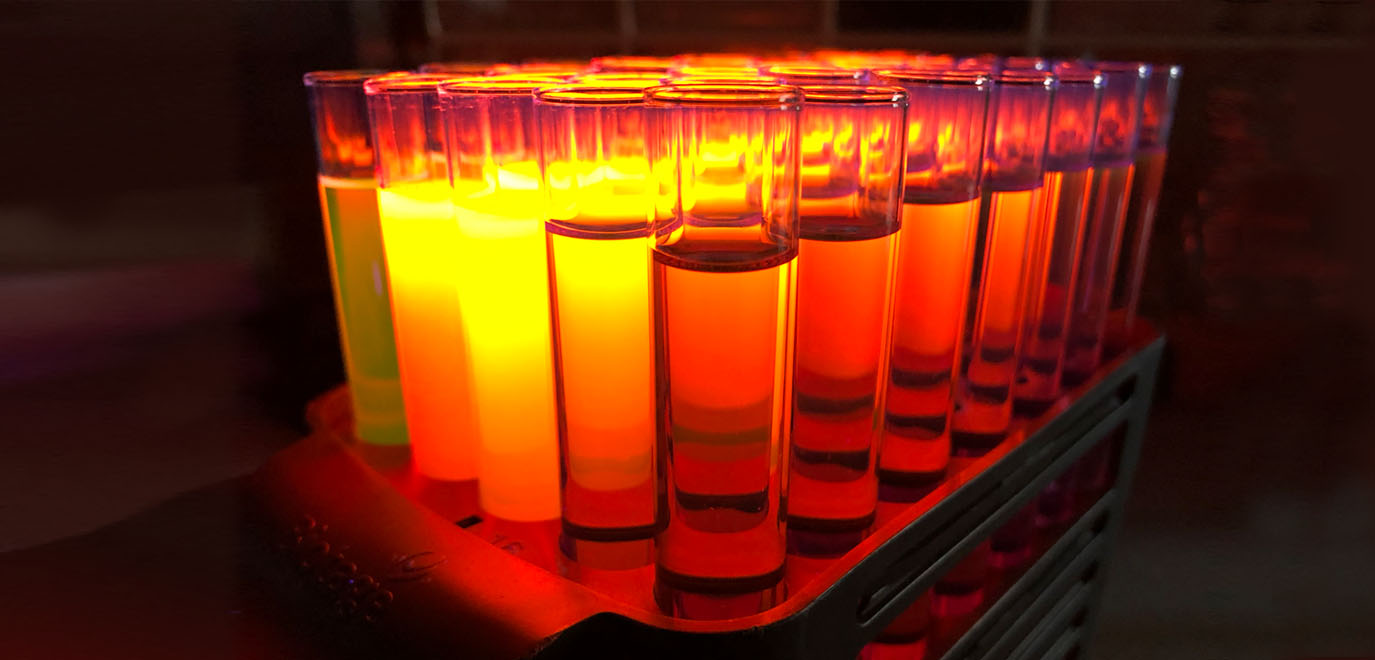
Test tubes of fluorescent dyes

Janelia Fluor® Dyes

The Lavis Lab improved upon small-molecule fluorophore synthesis to develop bright and photostable fluorescent dyes that enable imaging and tracking of individual molecules inside living cells.

area_landing_page_panel-jobs | block

Open Positions

area_landing_page_panel-full_bleed | block

“Janelia was founded on the idea that tool-builders should work alongside biologists to solve important scientific problems.”
-Luke Lavis

area_landing_page_panel-labs | block