Filter
Associated Lab
- Ahrens Lab (5) Apply Ahrens Lab filter
- Branson Lab (3) Apply Branson Lab filter
- Card Lab (1) Apply Card Lab filter
- Cardona Lab (1) Apply Cardona Lab filter
- Druckmann Lab (1) Apply Druckmann Lab filter
- Freeman Lab (4) Apply Freeman Lab filter
- Funke Lab (2) Apply Funke Lab filter
- Harris Lab (1) Apply Harris Lab filter
- Ji Lab (1) Apply Ji Lab filter
- Remove Keller Lab filter Keller Lab
- Lavis Lab (2) Apply Lavis Lab filter
- Liu (Zhe) Lab (1) Apply Liu (Zhe) Lab filter
- Looger Lab (5) Apply Looger Lab filter
- Pavlopoulos Lab (1) Apply Pavlopoulos Lab filter
- Tillberg Lab (1) Apply Tillberg Lab filter
- Turaga Lab (1) Apply Turaga Lab filter
Associated Project Team
Associated Support Team
Publication Date
- 2024 (1) Apply 2024 filter
- 2023 (1) Apply 2023 filter
- 2022 (1) Apply 2022 filter
- 2021 (2) Apply 2021 filter
- 2020 (5) Apply 2020 filter
- 2019 (6) Apply 2019 filter
- 2018 (6) Apply 2018 filter
- 2017 (2) Apply 2017 filter
- 2016 (6) Apply 2016 filter
- 2015 (7) Apply 2015 filter
- 2014 (7) Apply 2014 filter
- 2013 (9) Apply 2013 filter
- 2012 (3) Apply 2012 filter
- 2011 (2) Apply 2011 filter
- 2010 (2) Apply 2010 filter
60 Janelia Publications
Showing 11-20 of 60 results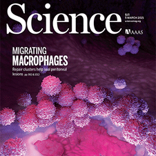
The mammalian heart is derived from multiple cell lineages; however, our understanding of when and how the diverse cardiac cell types arise is limited. We mapped the origin of the embryonic mouse heart at single-cell resolution using a combination of transcriptomic, imaging, and genetic lineage labeling approaches. This provided a transcriptional and anatomic definition of cardiac progenitor types. Furthermore, it revealed a cardiac progenitor pool that is anatomically and transcriptionally distinct from currently known cardiac progenitors. Besides contributing to cardiomyocytes, these cells also represent the earliest progenitor of the epicardium, a source of trophic factors and cells during cardiac development and injury. This study provides detailed insights into the formation of early cardiac cell types, with particular relevance to the development of cell-based cardiac regenerative therapies.

The origin of chordates has been debated for more than a century, with one key issue being the emergence of the notochord. In vertebrates, the notochord develops by convergence and extension of the chordamesoderm, a population of midline cells of unique molecular identity. We identify a population of mesodermal cells in a developing invertebrate, the marine annelid Platynereis dumerilii, that converges and extends toward the midline and expresses a notochord-specific combination of genes. These cells differentiate into a longitudinal muscle, the axochord, that is positioned between central nervous system and axial blood vessel and secretes a strong collagenous extracellular matrix. Ancestral state reconstruction suggests that contractile mesodermal midline cells existed in bilaterian ancestors. We propose that these cells, via vacuolization and stiffening, gave rise to the chordate notochord.
Calcium signaling has long been associated with key events of immunity, including chemotaxis, phagocytosis, and activation. However, imaging and manipulation of calcium flux in motile immune cells in live animals remain challenging. Using light-sheet microscopy for in vivo calcium imaging in zebrafish, we observe characteristic patterns of calcium flux triggered by distinct events, including phagocytosis of pathogenic bacteria and migration of neutrophils toward inflammatory stimuli. In contrast to findings from ex vivo studies, we observe enriched calcium influx at the leading edge of migrating neutrophils. To directly manipulate calcium dynamics in vivo, we have developed transgenic lines with cell-specific expression of the mammalian TRPV1 channel, enabling ligand-gated, reversible, and spatiotemporal control of calcium influx. We find that controlled calcium influx can function to help define the neutrophil's leading edge. Cell-specific TRPV1 expression may have broad utility for precise control of calcium dynamics in other immune cell types and organisms.
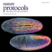
Light-sheet microscopy is a powerful method for imaging the development and function of complex biological systems at high spatiotemporal resolution and over long time scales. Such experiments typically generate terabytes of multidimensional image data, and thus they demand efficient computational solutions for data management, processing and analysis. We present protocols and software to tackle these steps, focusing on the imaging-based study of animal development. Our protocols facilitate (i) high-speed lossless data compression and content-based multiview image fusion optimized for multicore CPU architectures, reducing image data size 30–500-fold; (ii) automated large-scale cell tracking and segmentation; and (iii) visualization, editing and annotation of multiterabyte image data and cell-lineage reconstructions with tens of millions of data points. These software modules are open source. They provide high data throughput using a single computer workstation and are readily applicable to a wide spectrum of biological model systems.

Animal development is a complex and dynamic process orchestrated by exquisitely timed cell lineage commitment, divisions, migration, and morphological changes at the single-cell level. In the past decade, extensive genetic, stem cell, and genomic studies provided crucial insights into molecular underpinnings and the functional importance of genetic pathways governing various cellular differentiation processes. However, it is still largely unknown how the precise coordination of these pathways is achieved at the whole-organism level and how the highly regulated spatiotemporal choreography of development is established in turn. Here, we discuss the latest technological advances in imaging and single-cell genomics that hold great promise for advancing our understanding of this intricate process. We propose an integrated approach that combines such methods to quantitatively decipher in vivo cellular dynamic behaviors and their underlying molecular mechanisms at the systems level with single-cell, single-molecule resolution.
Understanding the diversification of mammalian cell lineages is an essential to embryonic development, organ regeneration and tissue engineering. Shortly after implantation in the uterus, the pluripotent cells of the mammalian epiblast generate the three germ layers: ectoderm, mesoderm and endoderm1. Although clonal analyses suggest early specification of epiblast cells towards particular cell lineages2–4, single-cell transcriptomes do not identify lineage-specific markers in the epiblast5–11 and thus, the molecular regulation of such specification remains unknow. Here, we studied the epigenetic landscape of single epiblast cells, which revealed lineage priming towards endoderm, ectoderm or mesoderm. Unexpectedly, epiblast cells with mesodermal priming show a strong signature for the endothelial/endocardial fate, suggesting early specification of this lineage aside from other mesoderm. Through clonal analysis and live imaging, we show that endothelial precursors show early lineage divergence from the rest of mesodermal derivatives. In particular, cardiomyocytes and endocardial cells show limited lineage relationship, despite being temporally and spatially co-recruited during gastrulation. Furthermore, analysing the live tracks of single cells through unsupervised classification of cell migratory activity, we found early behavioral divergence of endothelial precursors shortly after the onset of mesoderm migration towards the cardiogenic area. These results provide a new model for the phenotypically silent specification of mammalian cell lineages in pluripotent cells of the epiblast and modify current knowledge on the sequence and timing of cardiovascular lineages diversification.

Optical flow is a key method used for quantitative motion estimation of biological structures in light microscopy. It has also been used as a key module in segmentation and tracking systems and is considered a mature technology in the field of computer vision. However, most of the research focused on 2D natural images, which are small in size and rich in edges and texture information. In contrast, 3D time-lapse recordings of biological specimens comprise up to several terabytes of image data and often exhibit complex object dynamics as well as blurring due to the point-spread-function of the microscope. Thus, new approaches to optical flow are required to improve performance for such data. We solve optical flow in large 3D time-lapse microscopy datasets by defining a Markov random field (MRF) over super-voxels in the foreground and applying motion smoothness constraints between super-voxels instead of voxel-wise. This model is tailored to the specific characteristics of light microscopy datasets: super-voxels help registration in textureless areas, the MRF over super-voxels efficiently propagates motion information between neighboring cells and the background subtraction and super-voxels reduce the dimensionality of the problem by an order of magnitude. We validate our approach on large 3D time-lapse datasets of Drosophila and zebrafish development by analyzing cell motion patterns. We show that our approach is, on average, 10 x faster than commonly used optical flow implementations in the Insight Tool-Kit (ITK) and reduces the average flow end point error by 50% in regions with complex dynamic processes, such as cell divisions.
The comprehensive reconstruction of cell lineages in complex multicellular organisms is a central goal of developmental biology. We present an open-source computational framework for the segmentation and tracking of cell nuclei with high accuracy and speed. We demonstrate its (i) generality by reconstructing cell lineages in four-dimensional, terabyte-sized image data sets of fruit fly, zebrafish and mouse embryos acquired with three types of fluorescence microscopes, (ii) scalability by analyzing advanced stages of development with up to 20,000 cells per time point at 26,000 cells min(-1) on a single computer workstation and (iii) ease of use by adjusting only two parameters across all data sets and providing visualization and editing tools for efficient data curation. Our approach achieves on average 97.0% linkage accuracy across all species and imaging modalities. Using our system, we performed the first cell lineage reconstruction of early Drosophila melanogaster nervous system development, revealing neuroblast dynamics throughout an entire embryo.
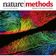
Recording light-microscopy images of large, nontransparent specimens, such as developing multicellular organisms, is complicated by decreased contrast resulting from light scattering. Early zebrafish development can be captured by standard light-sheet microscopy, but new imaging strategies are required to obtain high-quality data of late development or of less transparent organisms. We combined digital scanned laser light-sheet fluorescence microscopy with incoherent structured-illumination microscopy (DSLM-SI) and created structured-illumination patterns with continuously adjustable frequencies. Our method discriminates the specimen-related scattered background from signal fluorescence, thereby removing out-of-focus light and optimizing the contrast of in-focus structures. DSLM-SI provides rapid control of the illumination pattern, exceptional imaging quality, and high imaging speeds. We performed long-term imaging of zebrafish development for 58 h and fast multiple-view imaging of early Drosophila melanogaster development. We reconstructed cell positions over time from the Drosophila DSLM-SI data and created a fly digital embryo.
Histone post-translational modifications are key gene expression regulators, but their rapid dynamics during development remain difficult to capture. We applied a Fab-based live endogenous modification labeling technique to monitor the changes in histone modification levels during zygotic genome activation (ZGA) in living zebrafish embryos. Among various histone modifications, H3 Lys27 acetylation (H3K27ac) exhibited most drastic changes, accumulating in two nuclear foci in the 64- to 1k-cell-stage embryos. The elongating form of RNA polymerase II, which is phosphorylated at Ser2 in heptad repeats within the C-terminal domain (RNAP2 Ser2ph), and miR-430 transcripts were also concentrated in foci closely associated with H3K27ac. When treated with α-amanitin to inhibit transcription or JQ-1 to inhibit binding of acetyl-reader proteins, H3K27ac foci still appeared but RNAP2 Ser2ph and miR-430 morpholino were not concentrated in foci, suggesting that H3K27ac precedes active transcription during ZGA. We anticipate that the method presented here could be applied to a variety of developmental processes in any model and non-model organisms.